Search

Bienvenido a Canon Medical Academy

Su centro de acceso a contenido educativo avanzado y cursos de vanguardia impartidos por una red de expertos clínicos. Manténgase al día con los últimos avances en atención médica y maximice su contribución a la atención al paciente, gane confianza y mejore la eficiencia para aprovechar al máximo el potencial de su equipo Canon Medical.

Instalaciones de formación

Experimente un aprendizaje único a través de instalaciones de entrenamiento con equipos de última generación desarrollados para usted.

Cleveland, OH, USA
Image
Amstelveen, The Netherlands
Image
Nasu, Japan
Image

Educación Online

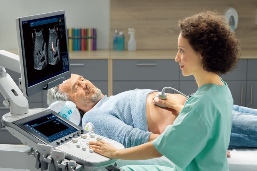
Mejore su crecimiento profesional con un amplio contenido a pedido, que incluye seminarios web, videos instructivos y una completa biblioteca de recursos.

Escalando la medicina deportiva con IA: movilidad de por vida y salud cardiovascular más allá del deporte


Fecha y hora:
Zona horaria 1: 26 de junio de 2025 | 19:00-20:10 (CEST) / 13:00-14:10 (EDT)
Zona horaria 2: 3 de julio de 2025 | 17:00-18:10 (SGT) / 19:00-20:10 (AEST)

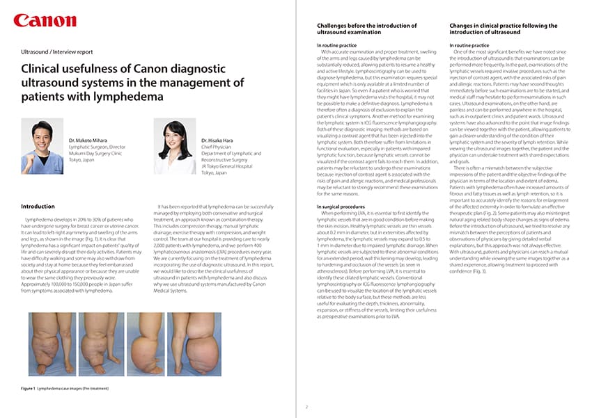
Esta sesión examina cómo la IA está escalando la medicina deportiva para promover la longevidad mediante el avance de la atención musculoesquelética y cardiovascular. Los temas incluyen el diagnóstico temprano basado en IA, la imagenología de precisión y la planificación personalizada del tratamiento y el entrenamiento. Destacaremos su papel en el apoyo a radiólogos, especialistas en ortopedia y cardiólogos en la estratificación del riesgo y la protección de la función musculoesquelética a lo largo de la vida, clave para mantener la movilidad, la salud cardiovascular y prolongar la esperanza de vida para todos.
Esperamos que nos acompañe.
Image

Other webinars

81 results. Try the Search box at the top, to filter these results.
Load more
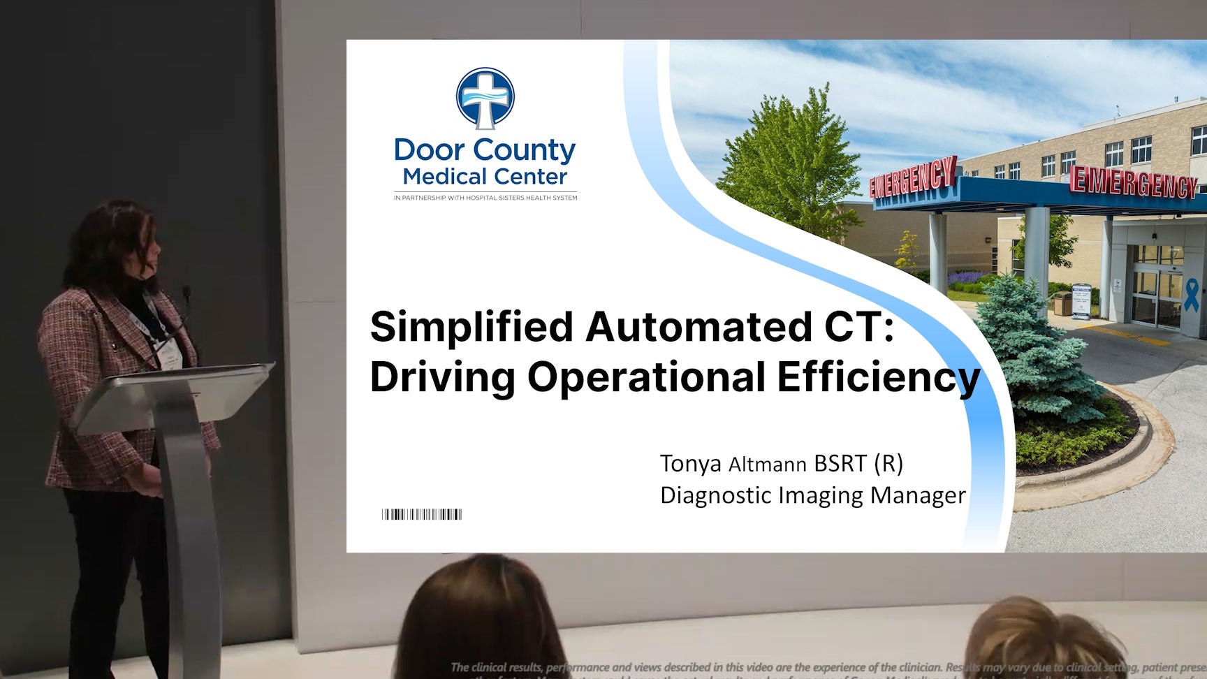
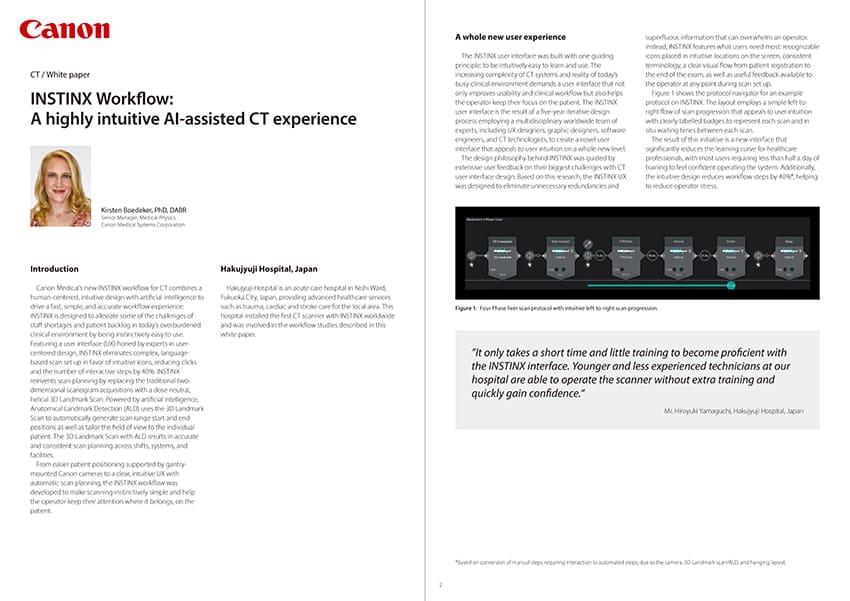
Simplified Automated CT: Driving Operational Efficiency

Radiology CT Educational lecture

Image

Simplified Automated CT: Enhanced Workflow

Radiology CT Educational lecture

Image

Moving Towards Autonomous CT

Radiology CT Educational lecture

Image

Advancements in CT chest imaging using the Canon platform

Lung | Radiology CT Educational lecture

Image

Next Generation Wide Area CT for Cardiothoracic Imaging: Faster, Sharper, Simpler

Cardiology | Pediatrics CT Educational lecture

Image

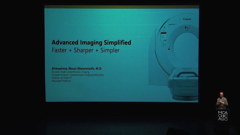
Advanced Imaging Simplified: Faster + Sharper + Simpler

Cardiology | Pediatrics CT Educational lecture

Image

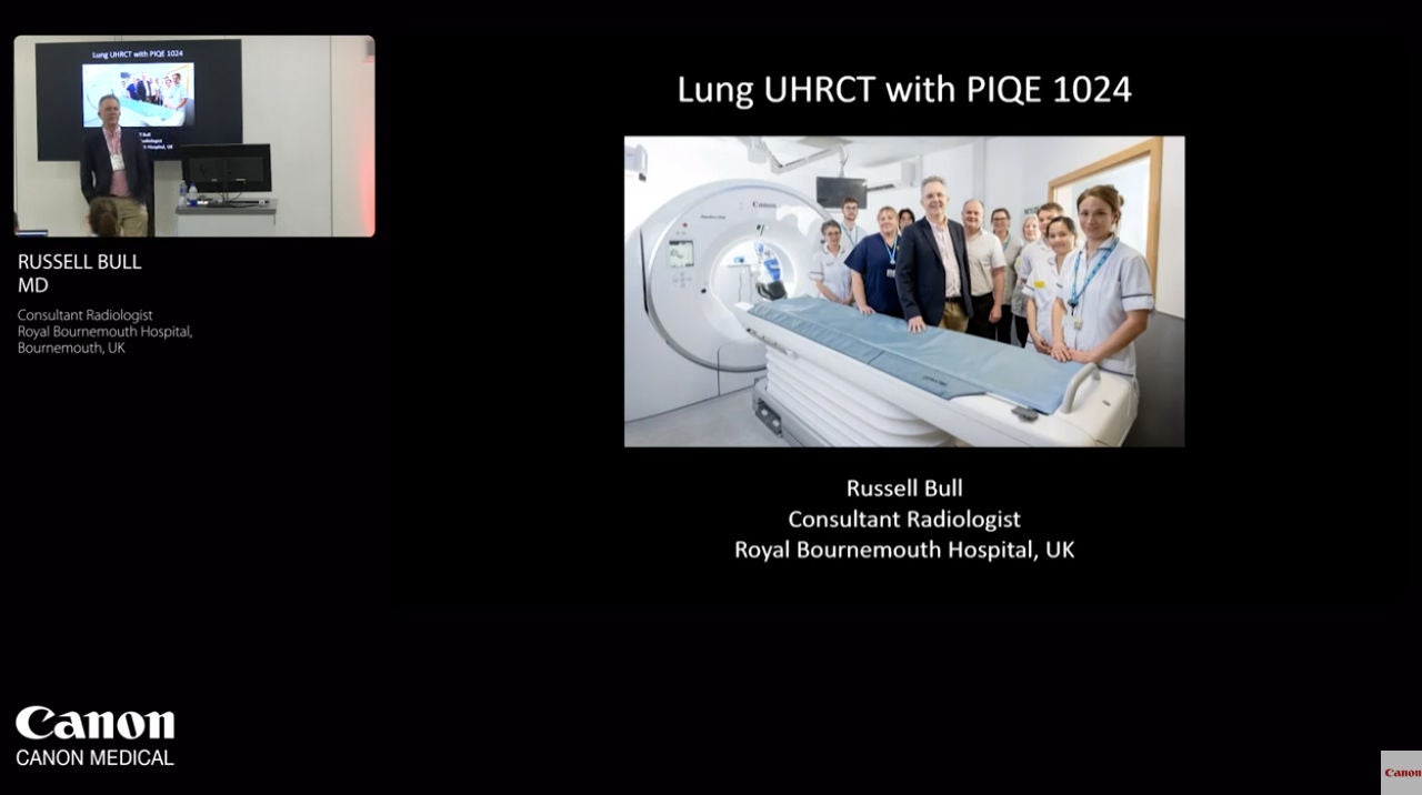
Ultra-High Resolution Lung CT: Pushing the Boundaries of Lung Imaging

Lung CT Educational lecture

Image

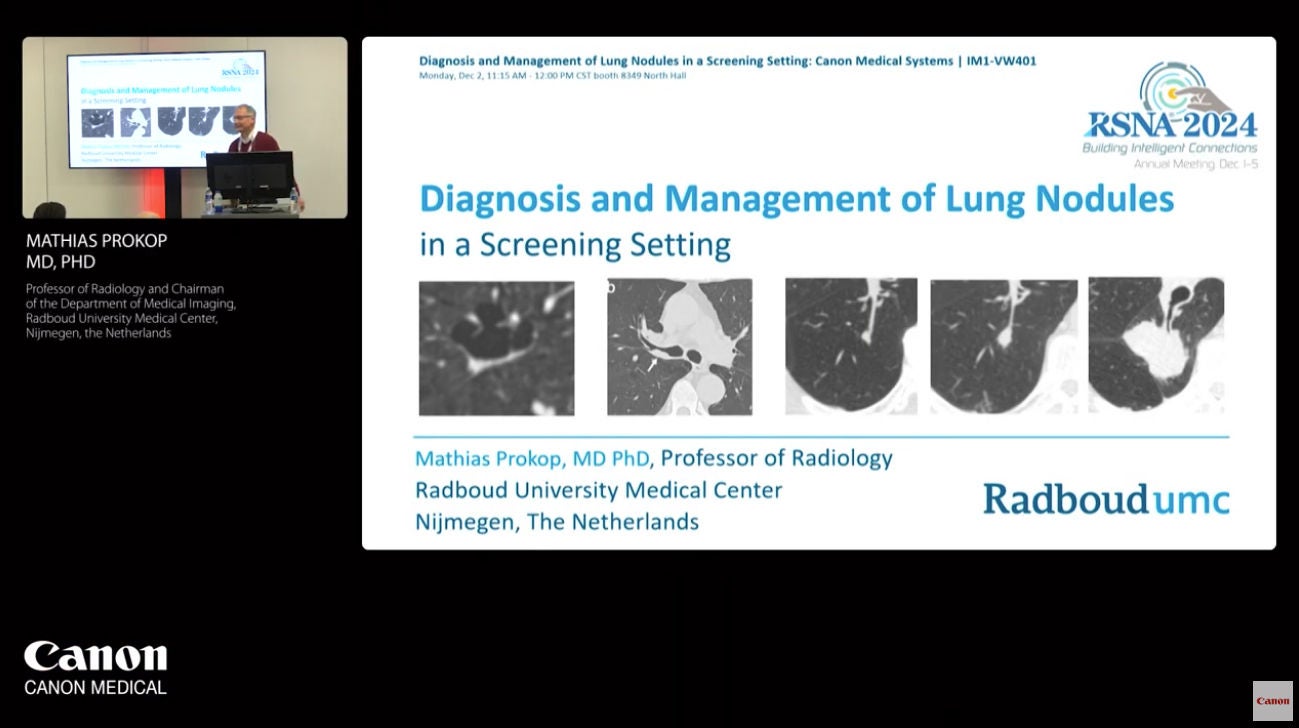
Diagnosis and Management of Lung Nodules in a Screening Setting

Lung | Lung Cancer Screening CT Educational lecture

Image

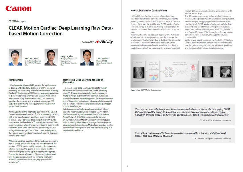
CLEAR Motion Cardiac: Deep Learning Raw Data-based Motion Correction

Gynecology | Women's Health Ultrasound Educational lecture

Image
Obstetrics | Women's Health Ultrasound Educational lecture
Image
Obstetrics | Women's Health Ultrasound Educational lecture
Image
Obstetrics | Women's Health Ultrasound Educational lecture
Image
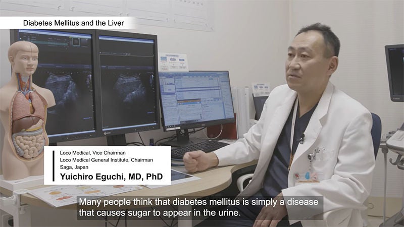
Liver | Radiology Ultrasound User experience video
Image
Liver | Radiology | Surgery Ultrasound User experience video
Image
Liver | Radiology Ultrasound Educational lecture
Image
General Imaging | Radiology | Urology Ultrasound User experience video
Image
General Imaging | Radiology | Urology Ultrasound User experience video
Image
Liver | Radiology Ultrasound Educational lecture
Image
Hepatology | Liver | Radiology Ultrasound Educational testimonial
Image
Radiology CT Video
Image
Cardiology | Radiology CT Video
Image
Radiology CT Video
Image
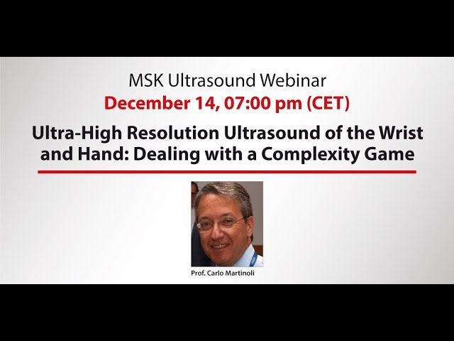
General Imaging | MSK Ultrasound Educational lecture
Image
General Imaging | MSK Ultrasound Educational lecture
Image
Liver | Radiology Ultrasound Educational testimonial
Image
Gastroenterology | Radiology Ultrasound User experience video
Image
Liver | Radiology Ultrasound Educational testimonial
Image
Liver | Radiology Ultrasound Educational lecture
Image
General Imaging | Radiology | Urology MRI | Ultrasound Educational lecture
Image
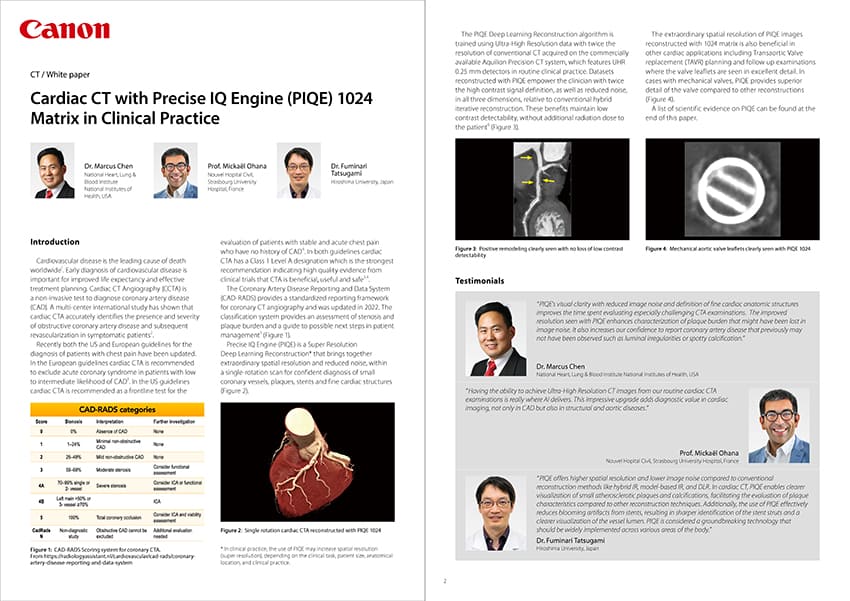
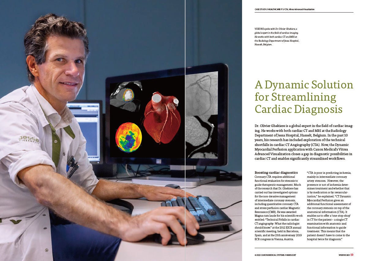
Cardiology | Radiology CT White paper
Image
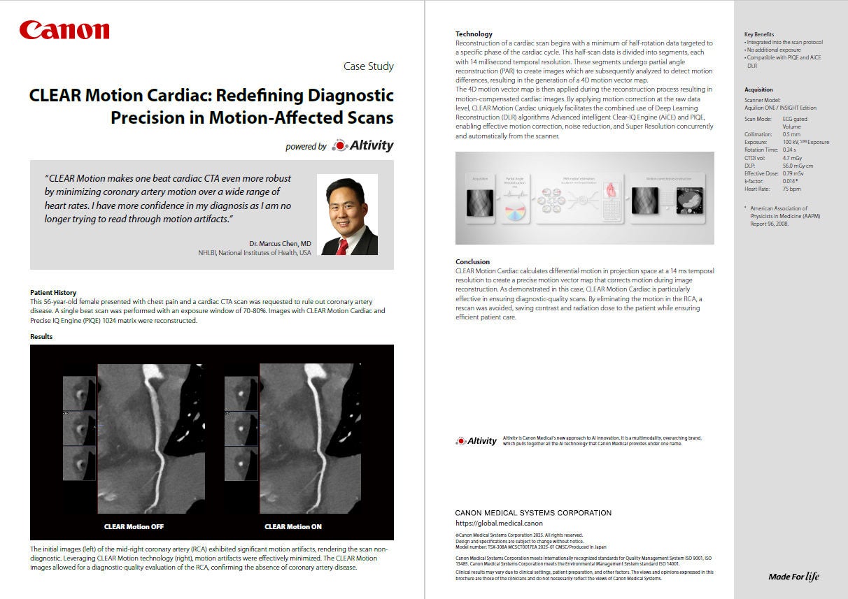
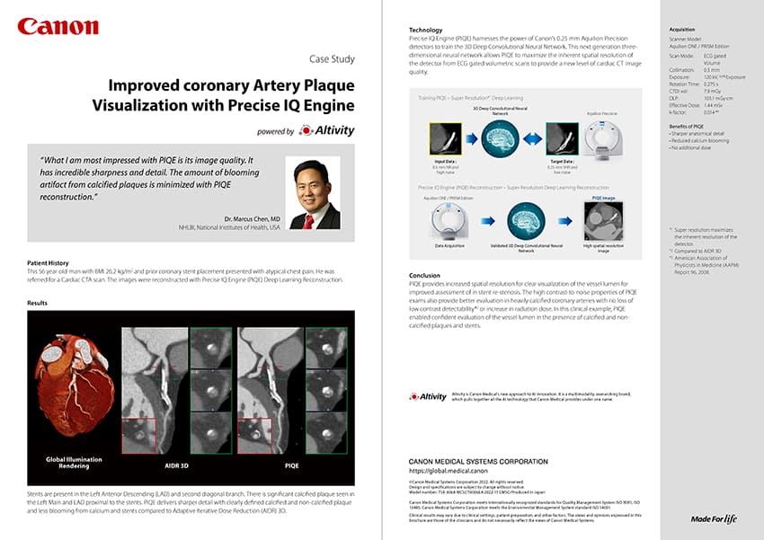
Cardiology | Radiology CT Case study
Image
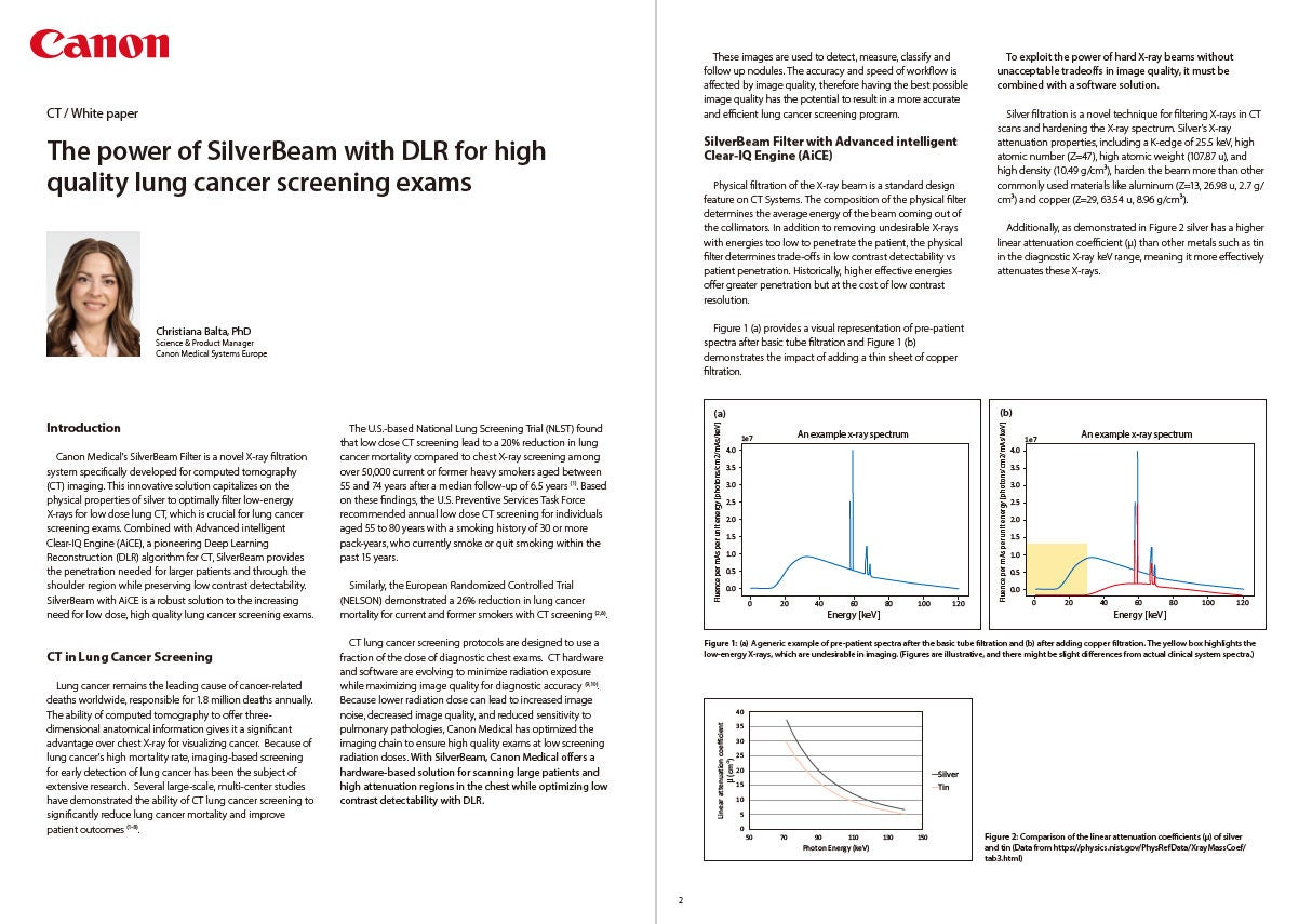
Lung Cancer Screening | Radiology CT White paper
Image
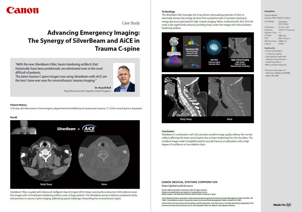
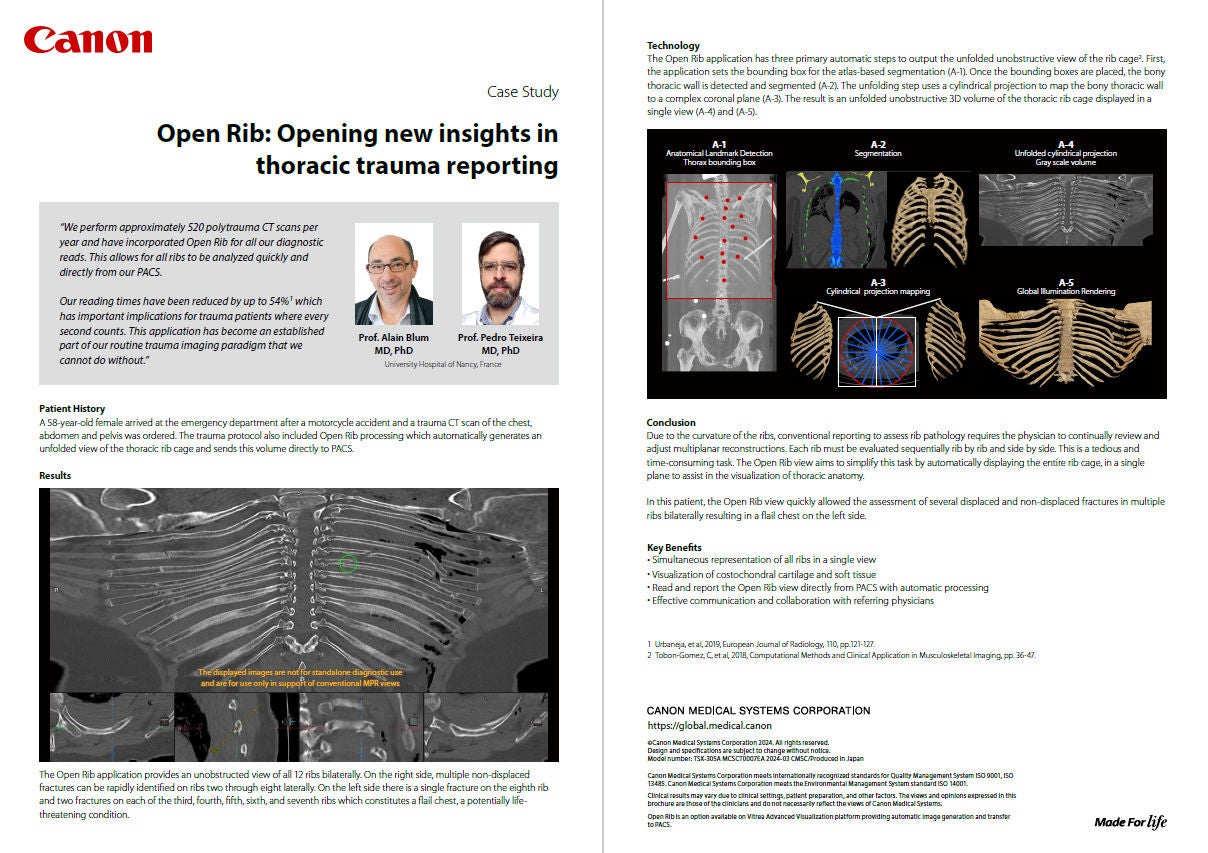
Emergency | MSK | Radiology CT Case study
Image
Radiology | Lung CT Case study
Image
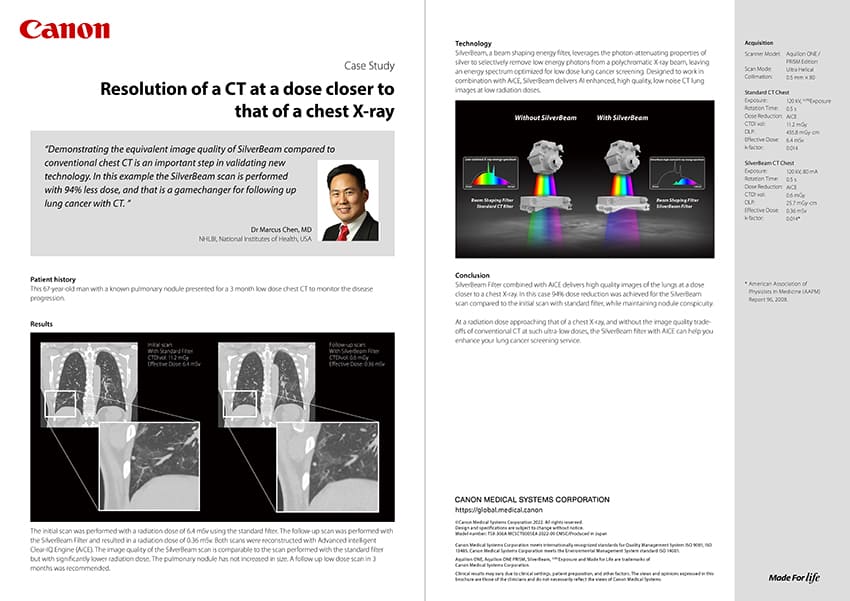
Lung Cancer Screening | Radiology CT | HIT Case study
Image
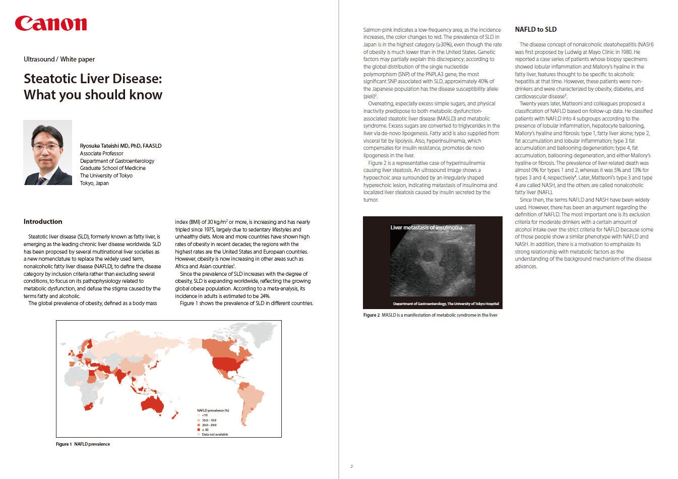
Radiology | Liver Ultrasound White paper
Image
Emergency | MSK CT | HIT Case study
Image
Liver | Radiology Ultrasound White paper
Image
Gastroenterology | Radiology Ultrasound White paper
Image
Radiology CT White paper
Image
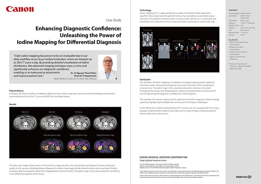
Liver | Radiology CT Case study
Image
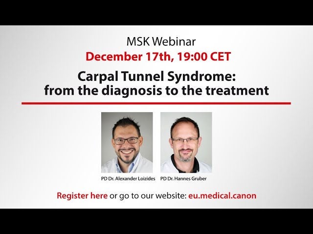
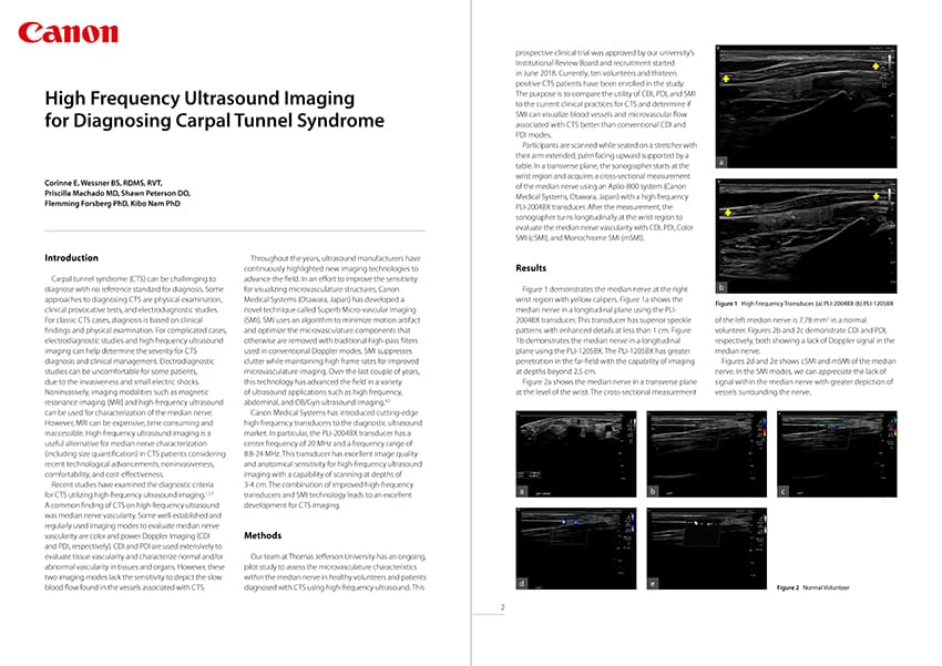
General Imaging | MSK Ultrasound White paper
Image
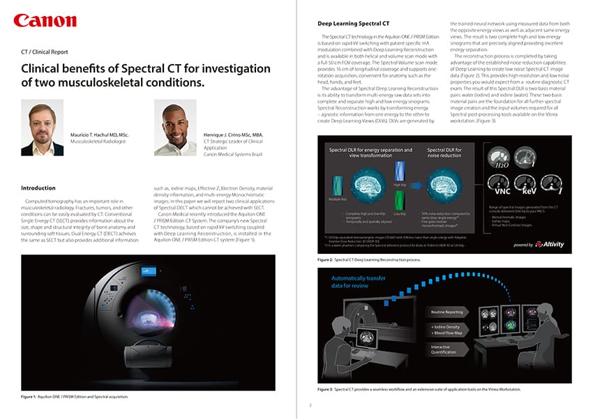
MSK | Radiology CT Clinical report
Image
Cardiology | Radiology CT White paper
Image
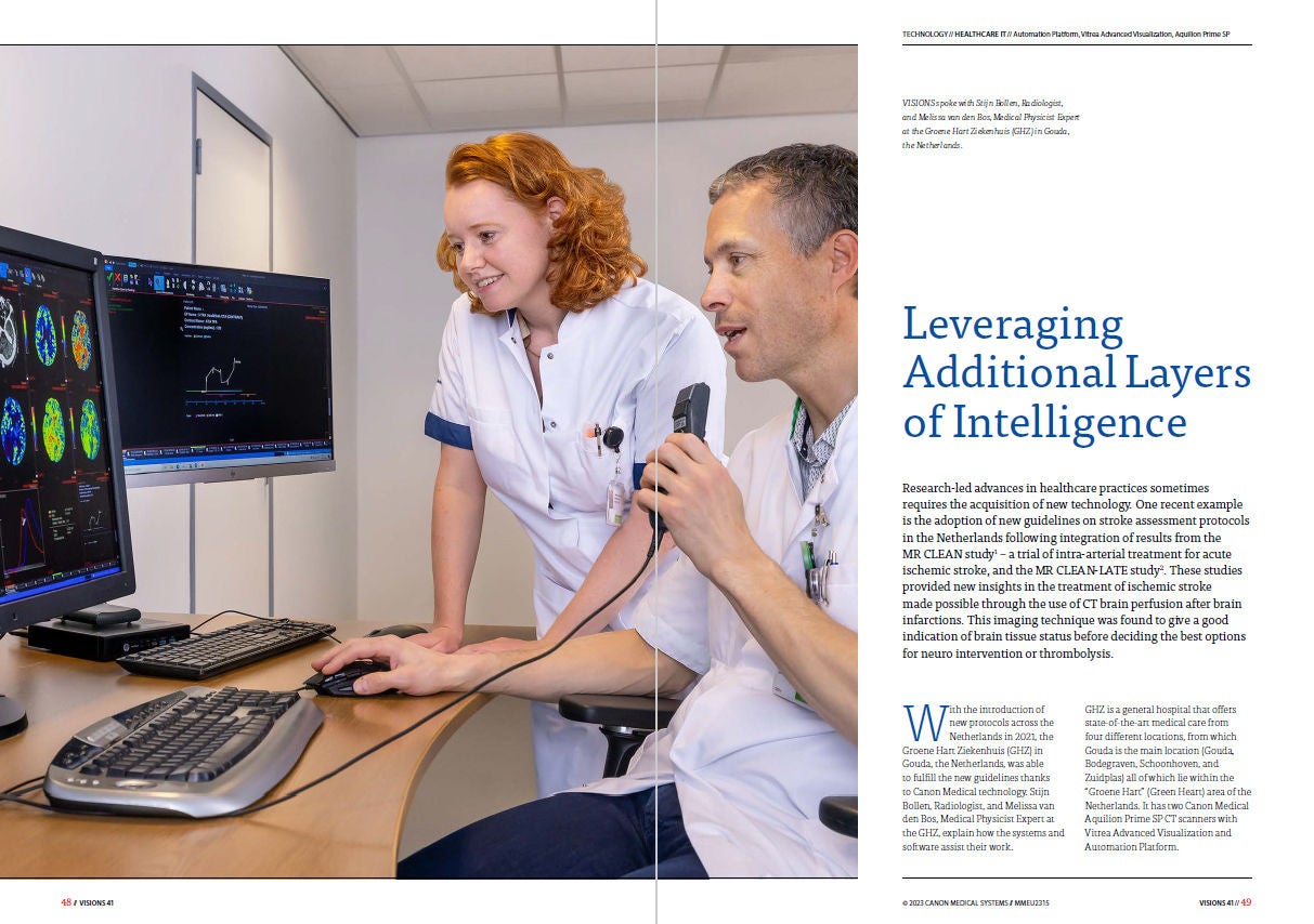
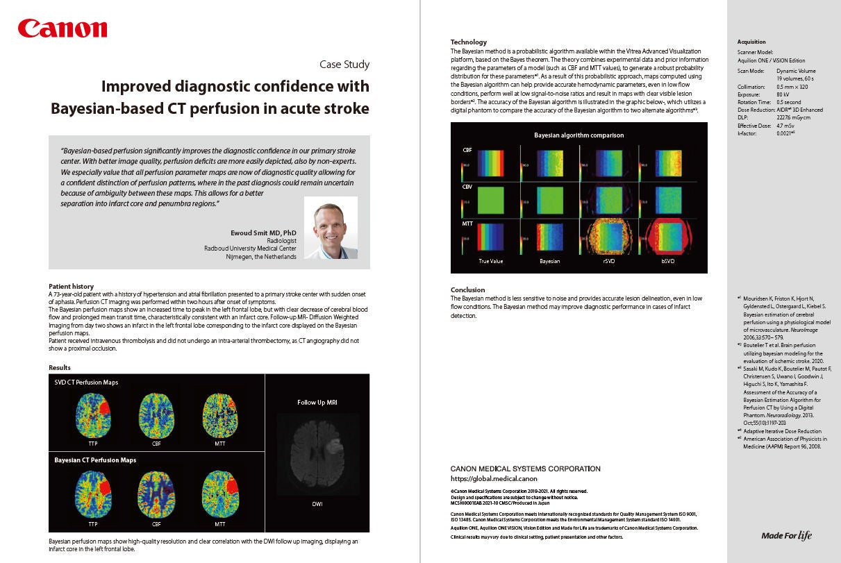
Stroke HIT Case study
Image
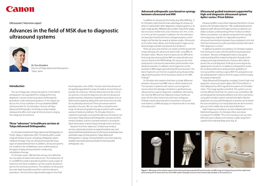
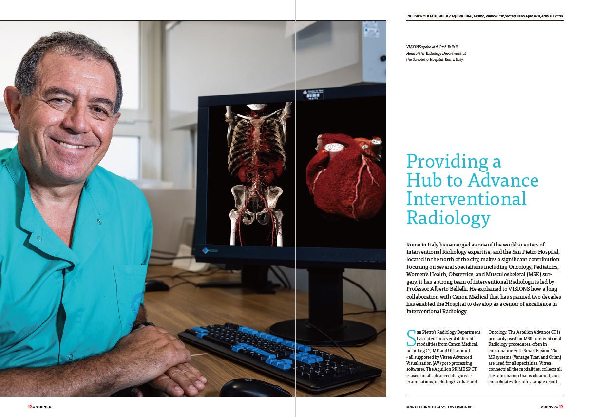
General Imaging | MSK Ultrasound Interview report
Image
General Imaging | Small parts Ultrasound Interview report
Image
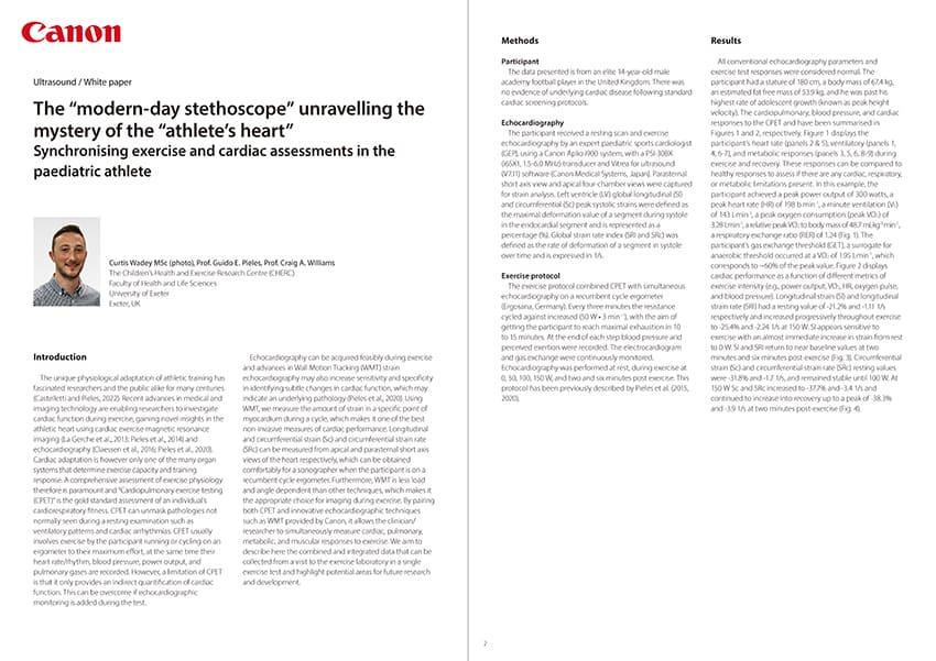
Cardiology | Pediatrics Ultrasound White paper
Image
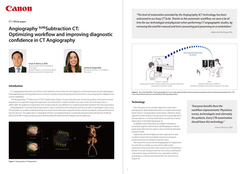
MSK | Radiology CT White paper
Image
General Imaging | MSK Ultrasound Interview report
Image
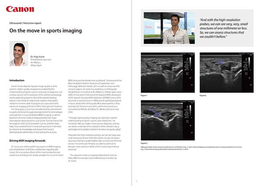
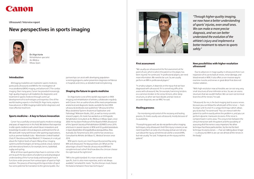
MSK | Sport Medicine Ultrasound Interview report
Image
Cardiology | Radiology CT Case study
Image
General Imaging | MSK Ultrasound Interview report
Image
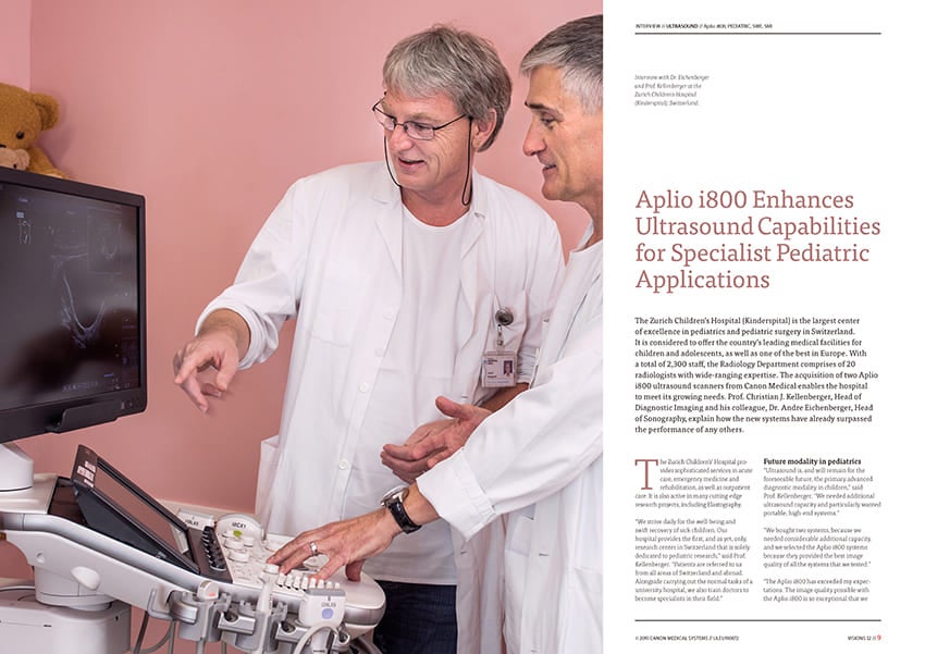
General Imaging | Pediatrics Ultrasound Interview report
Image
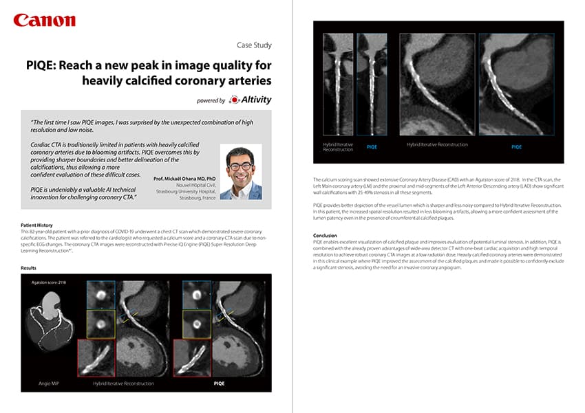
Cardiology | Radiology CT Case study
Image
Lung Cancer Screening | Radiology CT Case study
Image
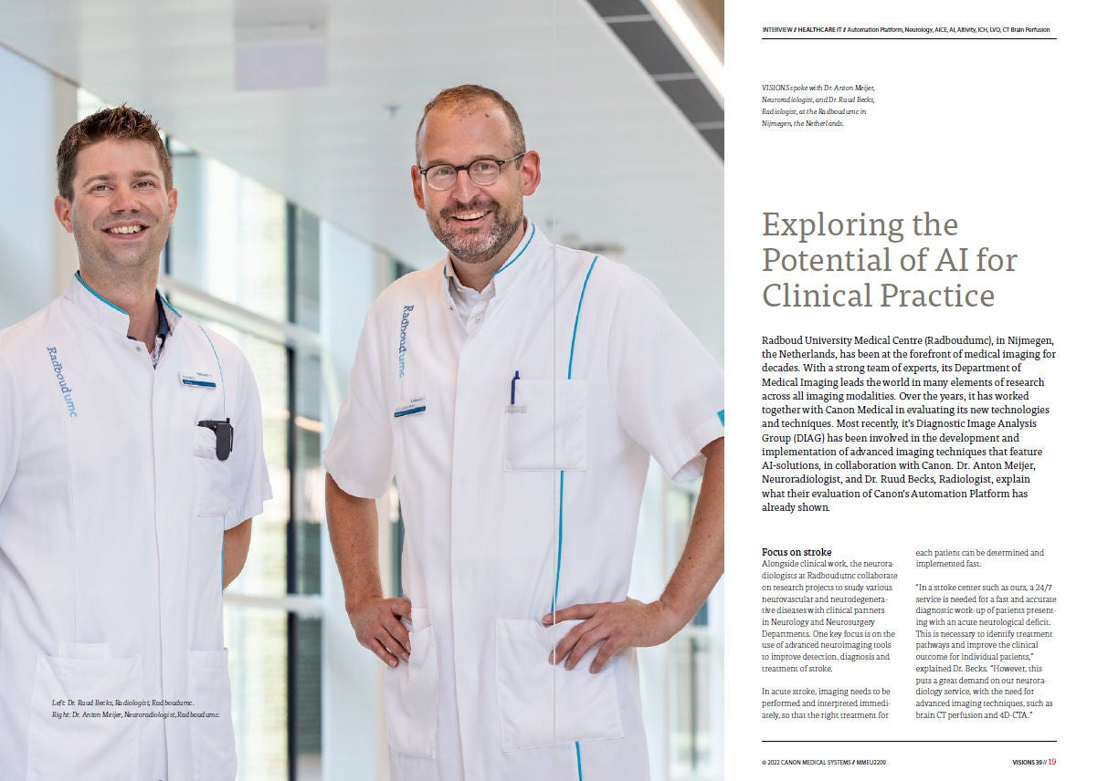
Stroke HIT Case study
Image
Stroke HIT Case study
Image
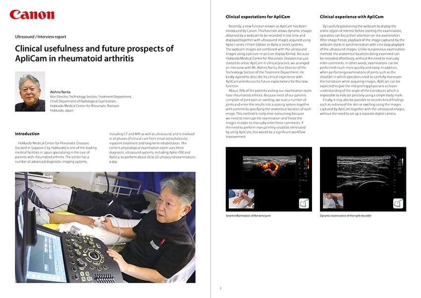
General Imaging | MSK Ultrasound Interview report
Image
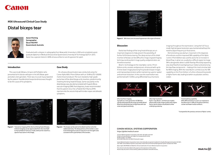
General Imaging | MSK Ultrasound Case study
Image
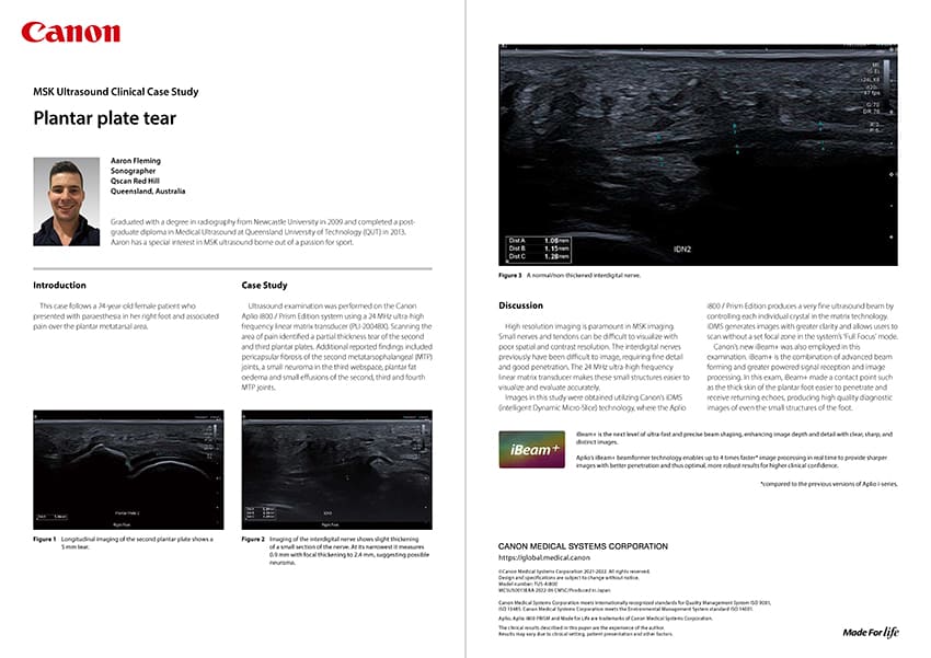
General Imaging | MSK Ultrasound Case study
Image
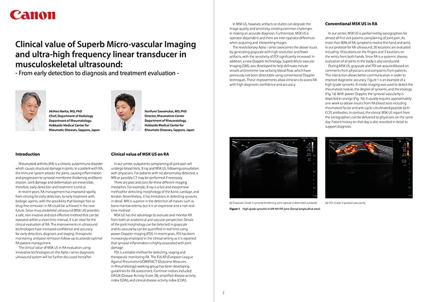
General Imaging | MSK Ultrasound White paper
Image
The global COVID-19 pandemic has greatly changed the environment surrounding Radiology. In last year’s RSNA2021, the theme of “Redefining Radiology” represented a defining frame for when we move past the pandemic. RSNA2021 highlighted workflow efficiency and patient-centeredness, while considering how to simultaneously maintain high-value imaging. MSK | Women's Health MR | Ultrasound Clinical report | Interview
Image
The Instituto Musculoesquelético Europeo (IMSKE; European Musculoskeletal Institute) is a new specialist hospital in Valencia, Spain. Opened in 2020, the pioneering facility is unique. It offers a wide spectrum of expertise in trauma care, including rheumatology, sport medicine, rehabilitation, physiotherapy within the context of plastic surgery, pediatric orthopedics, chiropody, nutrition, psychology, dermatology and neurology. The IMSKE has a multidisciplinary team of 70 muscu-loskeletal specialists and is fully equipped with Canon Medical imaging equipment. VISIONS explores how the institute is set to become the best traumatology hospital in Spain. Neuroradiology | Urology Ultrasound Interview | News
Image
VISIONS spoke with Chief Physician Prof. Lukas Lusuardi and Senior Physician Dr. Martin Drerup, University Department of Urology and Andrology at Salzburg University Hospital, Austria. Women's Health X-ray Technology
Image
The Handchirurgie Seefeld (hand surgery) clinic in Zürich, Switzerland, relies on high-tech medicine with the state-of-the-art Aplio i800 ultrasound diagnostic system of Canon Medical. Women's Health X-ray Technology
Image
VISIONS spoke with Prof. Kypros Nicolaides about using Canon's Aplio i-series systems in fetal medicine. Women's Health X-ray Technology
Image
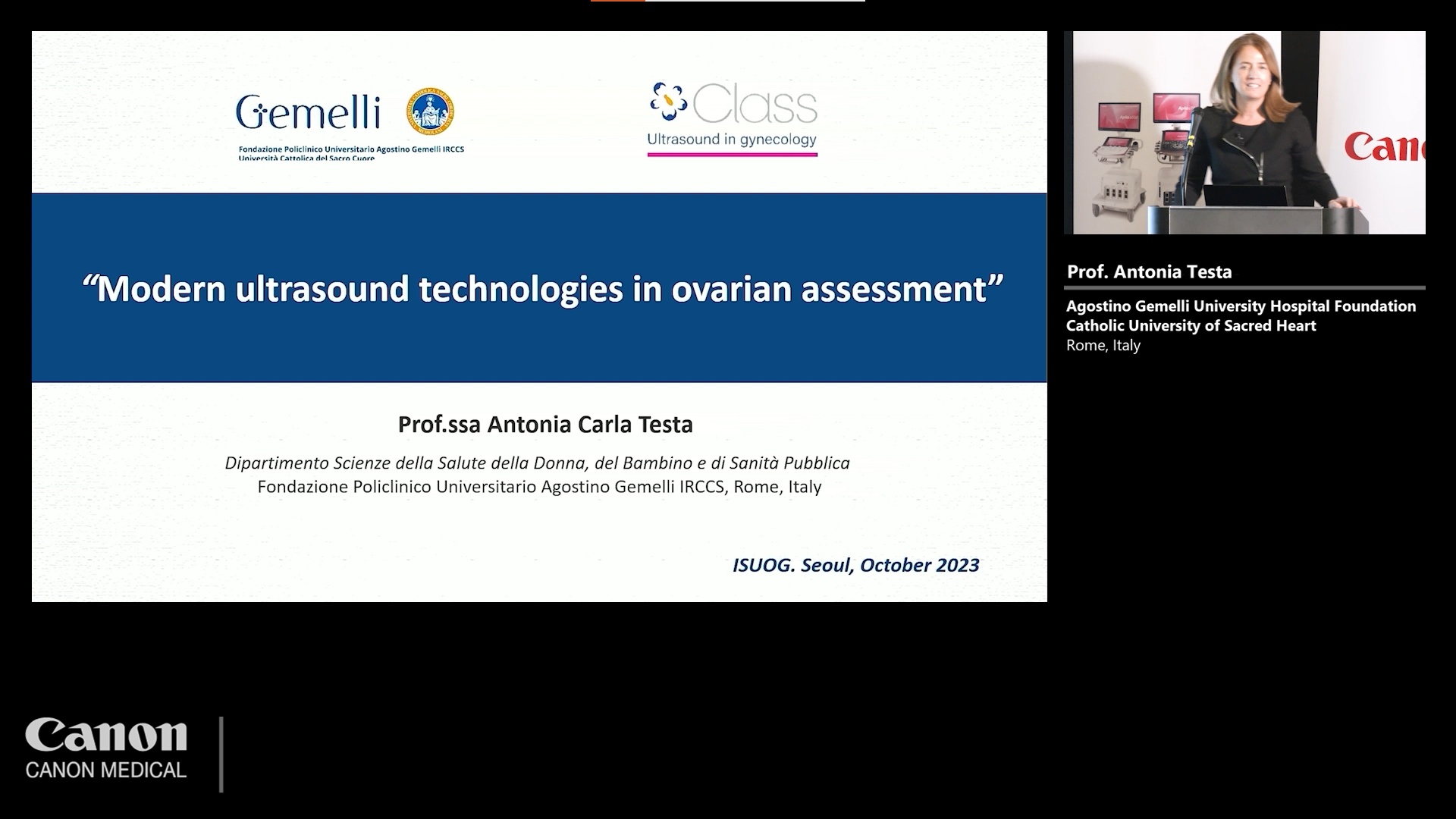
Professor Antonia Testa is one of Europe’s leading experts in gynecologic oncology and has conducted more than fifty research projects on gynecological cancer and chemotherapy protocols. She also founded a world-class school for ultrasound in gynecology in Rome, Italy, that has trained thousands of specialists since its inception. Professor Testa is also Board Member of the International Ovarian Tumor Analysis group (IOTA). Her overall work has contributed to improve early and confident diagnosis in gynecological disease. Professor Testa explained to VISIONS how important Canon Medical’s ultrasound systems are in daily clinical practice and research. Oncology | Urology Clinical application | Ultrasound Technology
Image
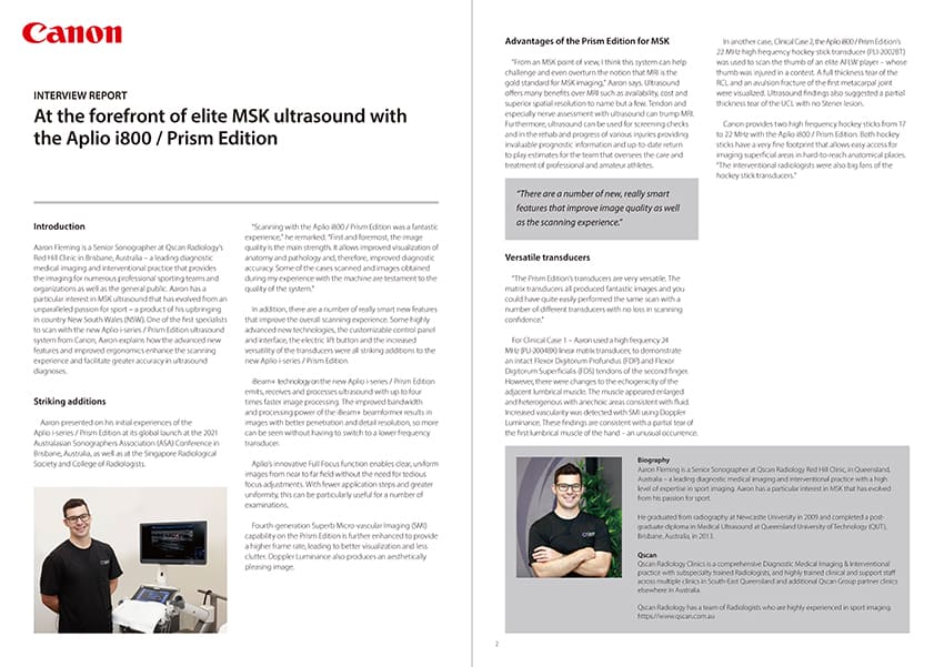
Aaron Fleming is a Senior Sonographer at Qscan Radiology’s Red Hill Clinic in Brisbane, Australia – a leading diagnostic medical imaging and interventional practice that provides the imaging for numerous professional sporting teams and organizations as well as the general public. Aaron has a particular interest in MSK ultrasound that has evolved from an unparalleled passion for sport – a product of his upbringing in country NSW. One of the first specialists to scan with the new Aplio i-series / Prism Edition ultrasound system from Canon Medical, Aaron explained to VISIONS how the advanced new features and improved ergonomics enhance the scanning experience and facilitate greater accuracy in ultrasound diagnoses. Women's Health X-ray Technology
Image
Cardiology HIT Case study
Image
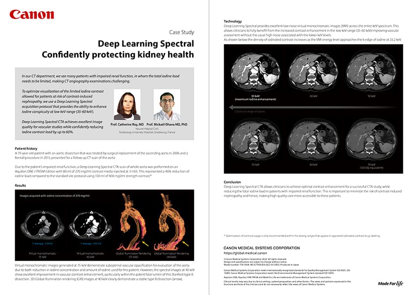
Kidney | Radiology CT Case study
Image
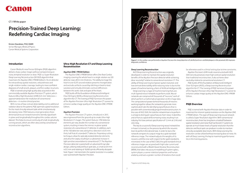
Cardiology | Radiology CT White paper
Image
Neurology HIT Case study
Image
MSK HIT Case study
Image
VISIONS spoke with Dr. Frédéric Alexis (Head of the Radiology department at Sainte Elisabeth Clinic, Namur, Belgium) about the experiences with Canon Medical's CT systems; the Aquilion Lightning SP, the Aquilion ONE / PRISM Edition, and the Aquilion Precision. Oncology Clinical application | CT | MR Interview | News
Image
Canon Medical and the Department of Radiology, Uppsala University Hospital, Sweden, will celebrate this November the ten-year anniversary collaborative partnership in our neurointervention labora-tory. The relationship between Canon Medical and our section has never been an ordinary suppli-er-customer relationship. These ten years have been marked by mutual efforts in the development of the biplane system and on work that has focused on an analysis of the unique functionalities of this system. The conclusions of our strategic discussions are implemented by Canon’s R & D teams. All challenges have been overcome thanks to energy, motivation and, first of all, the deep mutual respect which was established during the many years of our collaboration. The Alphenix Biplane system that was launched was partly the result of these mutual efforts. Cardiology MRI | Ultrasound Case study
Image
Pétur Hannesson and Steinuum Erla Thoracius, both Head of the Radiology department, and Kristín Rut Haraldóttir, responsible for the education and training of midwives in Ultrasound at Landspitali (National University Hospital) in Reykjavik, Iceland, share their experiences with Canon Medical’s equipment in this dedicated interview with VISIONS magazine. Neurology | Oncology HIT | Ultrasound News | Product
Image
One of the main challenges in MRI is finding the optimal balance of Signal to Noise Ratio (SNR) and Image Resolution such that detailed structures can be clearly visualized. While nearly every radiologist dreams of ultrahigh resolution, the reality remains that when spatial resolution is increased, the available signal per voxel decreases, leading to lower SNR. Image noise can then hinder visualization of critical features and introduce uncertainty and frustration into the diagnostic process. When noise becomes overly apparent, technologists have no choice but to manipulate parameters, commonly leading to both protocol inconsistency and prolongation of scan times. These undesired consequences negatively affect patient care and decrease productivity in the radiology department. Cerebrovascular Disease | MSK HIT | MR Technology
Image
Borderline ovary tumors usually affect young women and are symptom-free. This type of tumor is different from cancer. This article will help you to understand the differences and how to distinguish borderline lesions from other types of lesions. Borderline ovarian tumors are usually diagnosed at an early stage. The main therapy for these tumors is surgery. There is a small risk of tumor recurrence. Borderline ovarian tumors are lesions with certain abnormalities, shaped within the tissue covering the ovary. They are often discovered during an ultrasound exam or an infertility assessment, for example. Tumors of this type are not malignant and are usually treated by surgery. About 15 out of every 100 (15%) ovarian tumors are borderline tumors. They are also called "tumors with low malignant potential". Borderline tumors usually affect women between 20 and 40 years old. There are two main types of borderline ovarian tumors: serous and mucinous. Serous borderline ovarian tumors are the most common. They represent about 65% of cases of borderline ovarian tumors. Borderline ovarian tumors are different from cancer (Figure 1) because they do not grow in ovarian support tissue (the stroma). They tend to grow slowly and more controlled than cancer cells. Occasionally, some abnormal cells come off the tumor and settle elsewhere in the body, usually in the abdomen. Very rarely, these cells begin to develop in the underlying tissue (peritoneal carcinomatosis). Women's Health CT | MR Product | Technology
Image
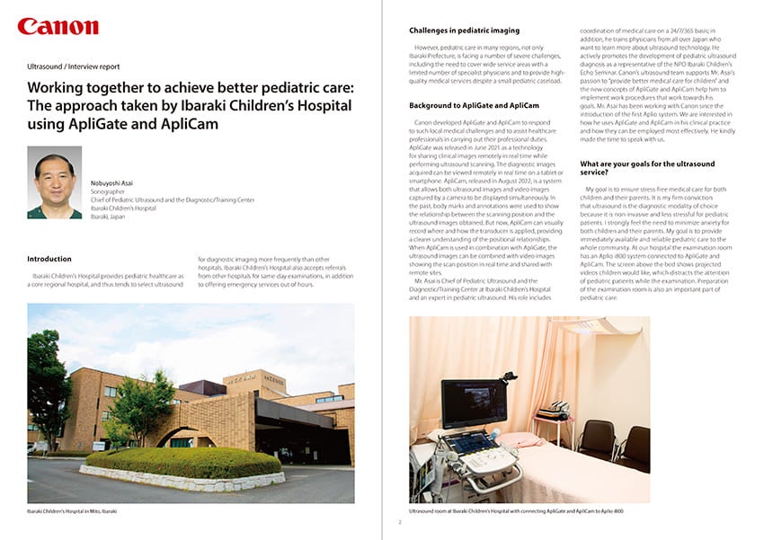
General Imaging | Pediatrics | Radiology Ultrasound Interview report
Image
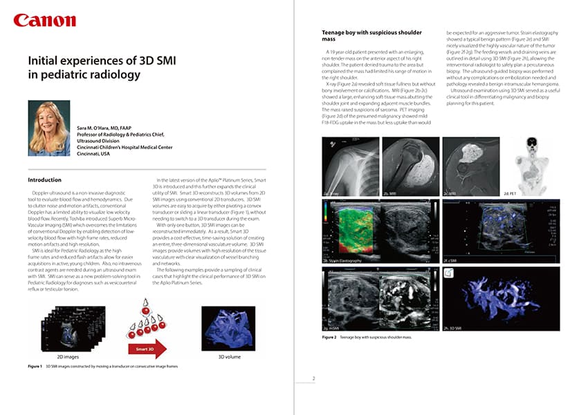
General Imaging | Pediatrics Ultrasound White paper
Image
Due to medical device regulatory reasons, not all products/service displayed on this Canon Medical Systems Argentina webpage are available in all countries, regions or markets. Future availability of the products/service cannot also be guaranteed. Please contact your local Canon Medical Systems representative for further details.

Image