

Highlights of some key findings
At Canon Medical, we are collaborating with researchers worldwide to develop innovative stroke solutions. Explore some of our key findings here.
Ultrasound
What is the role of Superb Micro-vascular Imaging (SMI) in the detection of carotid plaque neovascularization?
Intraplaque neovascularization (IPN) is a key feature of plaque instability but its detection is traditionally difficult with B-mode and Doppler Imaging.
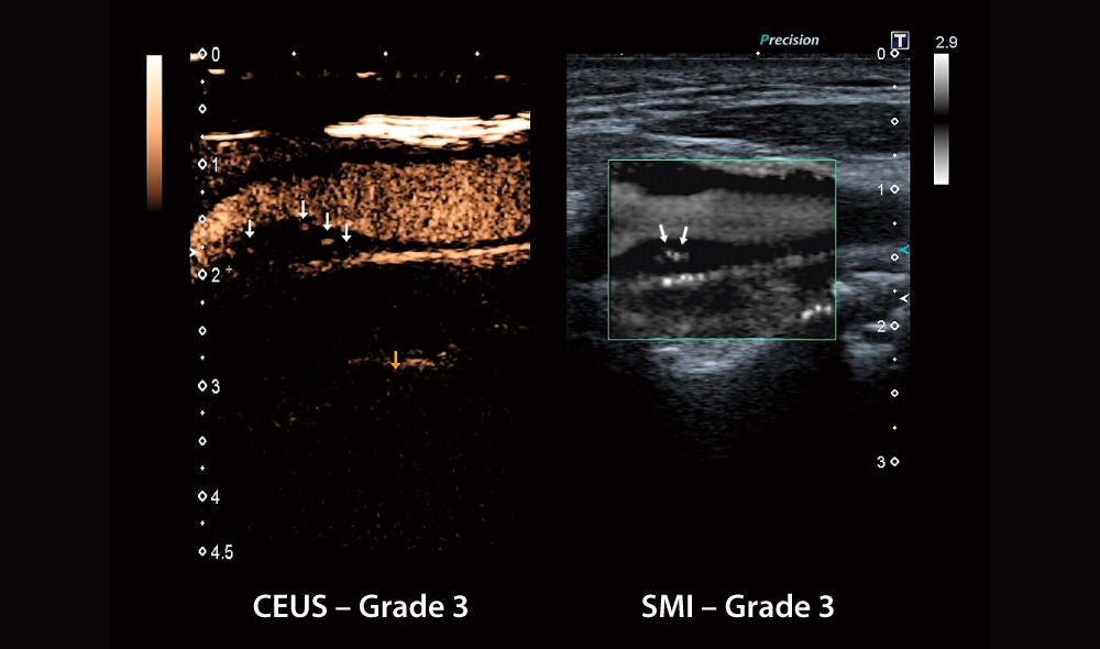 Case example of an asymptomatic patient with more than 70% carotid stenosis (predominantly hypoechoic plaque) in the right internal carotid artery. On CEUS, bubbles moving into the plaque core can be observed. Similarly, intraplaque microvascular flow signals moving towards the plaque core can be observed on SMI as well.
- SMI performed in 31 patients with internal carotid stenosis ≥50% and compared to CEUS and histology
- Very strong relationship between SMI and CEUS (correlation r=0.911, P<0.001)
- Plaques with higher IPN grades on SMI also showed significantly more neovessels on histology
Conclusion: “These findings demonstrate that SMI can detect neovascularization with accuracy comparable to CEUS, suggesting SMI as a promising noninvasive alternative to CEUS.”
Reference:
Zamani et al. | Carotid Plaque Neovascularization Detected With Superb Microvascular Imaging Ultrasound Without Using Contrast Media | Stroke (2019)
https://pubmed.ncbi.nlm.nih.gov/31510899/
CT x HIT
How does whole brain (16 cm) perfusion vs. traditional brain (4 cm) perfusion support decision making?
With a scanner that acquires less than the entire brain in a rotation, the user has to make sacrifices in either coverage or accuracy. This study compared the impact of performing CT perfusion with whole brain (16 cm) coverage versus traditional (4 cm) coverage in the assessment of cerebral ischemia.
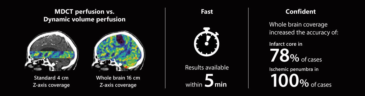
Conclusion: “The increased field of view better defined the true extent of the identified infarct core and ischemic penumbra. It also showed other areas of infarction that were not identified on the 4 cm Z-axis.”
Reference:
Page et al. | Comparison of 4 cm Z-axis and 16 cm Z-axis multidetector CT perfusion | European radiology (2010)
https://pubmed.ncbi.nlm.nih.gov/20013273/
Healthcare IT
What is the performance of Automation Platform (AP) in the detection of large vessel occlusion (LVO)?
303 consecutive patients with acute ischemic stroke symptoms.
Time from pushing case to receiving LVO results is < 70s.
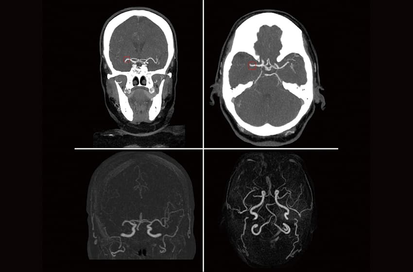
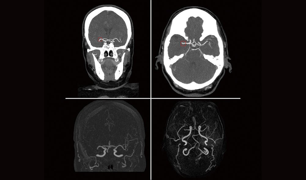 Correctly predicted large vessel occlusion (LVO) in case with right middle cerebral artery (MCA) occlusion. Top row shows coronal and axial views of the correctly labeled LVO, as indicated by the red box*. Bottom row shows the same case with 2D MIP subtraction and 3D MIP subtraction (right) where you can visualize the lack of contrast distal to the occlusion. * Not available in all geographies.
All cases; n=303:
Accuracy 81%
Sensitivity 73%
Specificity 98%
Internal carotid artery (ICA); n=160:
Accuracy 95%
Sensitivity 90%
Specificity 98%
Conclusion: “Canon’s Automation Platform Stroke CT LVO application was able to accurately identify ICA and M1 MCA occlusions in addition to almost perfectly assessing when an LVO was not present.”
Reference:
Rava et al. | Validation of an Artificial Intelligence Driven Large Vessel Occlusion Detection Algorithm for Acute Ischemic Stroke Patients | The Neuroradiology Journal (2021)
https://pubmed.ncbi.nlm.nih.gov/33657922/
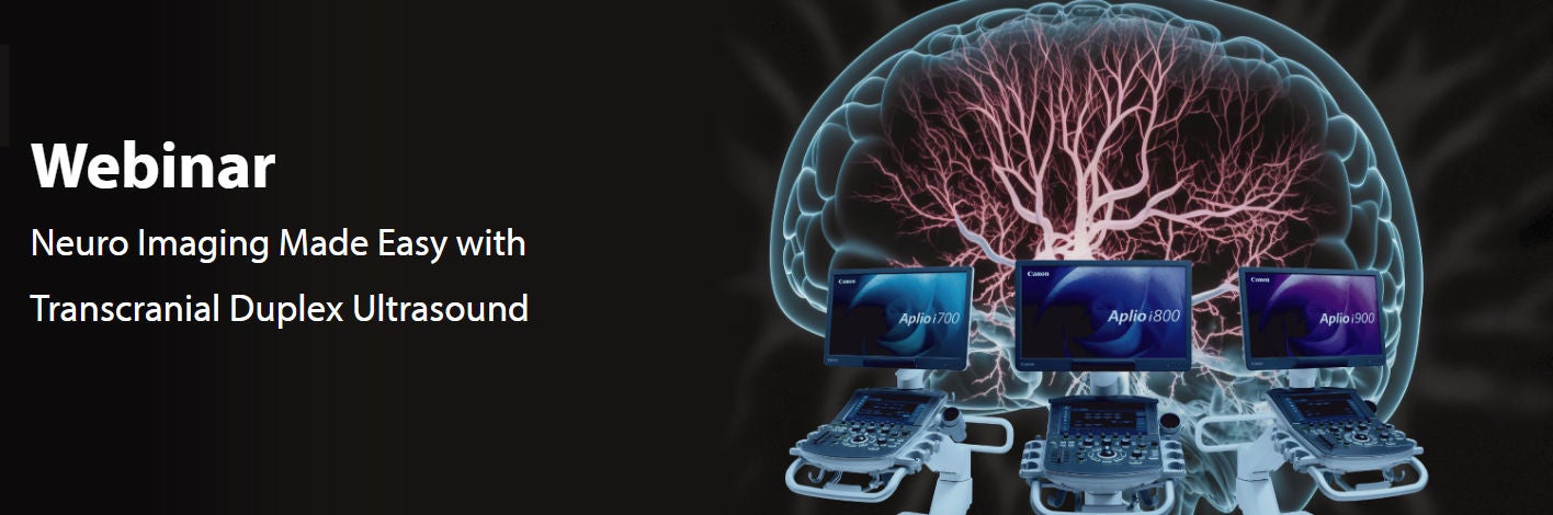
Online webinars
Watch our latest online webinars, where leading experts from around the world come to share discoveries and insights from research and clinical practice in stroke care.


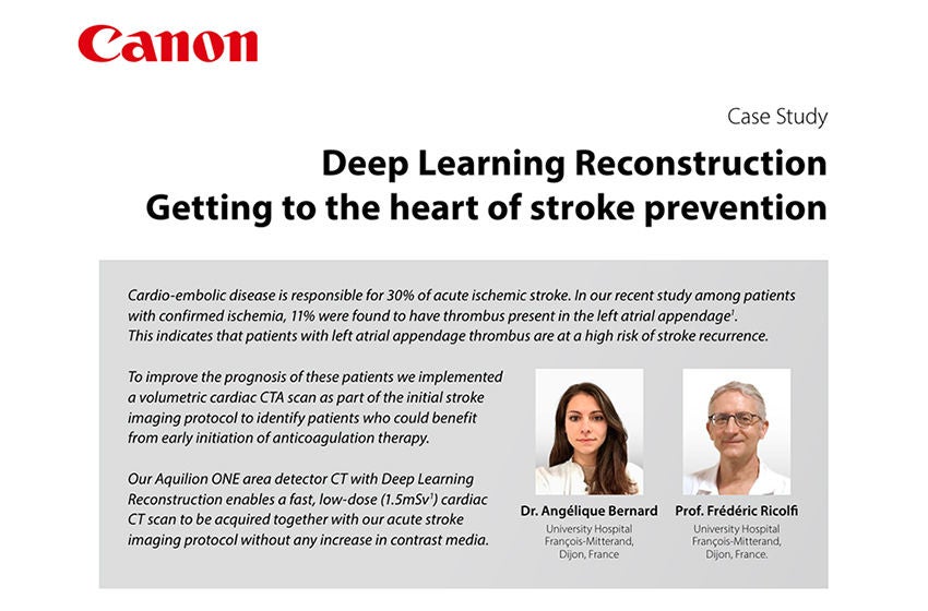
White papers and case studies
Find our latest white papers and case studies on stroke solutions using Canon technologies here.
Risk Stratification
Triage & Diagnosis



Treatment
Scientific papers
Find our latest scientific evidence on stroke solutions using Canon technologies here.
Guo et al. | The Value of Superb Microvascular Imaging and Contrast- enhanced Ultrasound for the Evaluation of Neovascularization in Carotid Artery Plaques | Academic radiology (2023)
https://pubmed.ncbi.nlm.nih.gov/36123231/
Gao et al. | Assessment of Carotid Body Tumors by Superb Microvascular Imaging of Feeding Arteries During Preoperative Evaluation | Frontiers in surgery (2022)
https://pubmed.ncbi.nlm.nih.gov/35558392/
Hagiwara et al. | Carotid Ultrasound Using Superb Microvascular Imaging to Identify Patients Developing In- Stent Restenosis After CAS | Journal of stroke and cerebrovascular diseases : the official journal of National Stroke Association (2022)
https://pubmed.ncbi.nlm.nih.gov/35797762/
Li, Y et al. | Advance ultrasound techniques for the assessment of plaque vulnerability in symptomatic and asymptomatic carotid stenosis: a multimodal ultrasound study | Cardiovascular diagnosis and therapy (2021)
https://pubmed.ncbi.nlm.nih.gov/33708475/
Meng et al. | Assessment of neovascularization of carotid artery atherosclerotic plaques using superb microvascular imaging: a comparison with contrast- enhanced ultrasound imaging and histology | Quantitative imaging in medicine and surgery (2021)
https://pubmed.ncbi.nlm.nih.gov/33936978/
Chiba et al. | Superb Microvascular Imaging Ultrasound for Cervical Carotid Artery Stenosis for Prediction of the Development of Microembolic Signals on Transcranial Doppler during Carotid Exposure in Endarterectomy | Cerebrovascular diseases extra (2021)
https://pubmed.ncbi.nlm.nih.gov/34034253/
Song et al. | Detection of Carotid Atherosclerotic Intraplaque Neovascularization Using Superb Microvascular Imaging: A Meta- Analysis | Journal of ultrasound in medicine : official journal of the American Institute of Ultrasound in Medicine (2021)
https://pubmed.ncbi.nlm.nih.gov/33587302/
Jin et al. | Differential value of intima thickness in ischaemic stroke due to large- artery atherosclerosis and small- vessel occlusion | Journal of cellular and molecular medicine (2021)
https://pubmed.ncbi.nlm.nih.gov/34459107/
Xu et al. | The Diagnostic Value of Radial and Carotid Intima Thickness Measured by High- Resolution Ultrasound for Ischemic Stroke | Journal of the American Society of Echocardiography : official publication of the American Society of Echocardiography (2021)
https://pubmed.ncbi.nlm.nih.gov/33121857/
Yang et al. | Consistency of superb microvascular imaging and contrast- enhanced ultrasonography in detection of intraplaque neovascularization: A meta- analysis | PloS one (2020)
https://pubmed.ncbi.nlm.nih.gov/32730304/
Chen et al. | Neovascularization in carotid atherosclerotic plaques can be effectively evaluated by superb microvascular imaging (SMI): Initial experience | Vascular medicine (London, England) (2020)
https://pubmed.ncbi.nlm.nih.gov/32303154/
Kaszczewski et al. | Volumetric Carotid Flow Characteristics in Doppler Ultrasonography in Healthy Population Over 65 Years Old | Journal of clinical medicine (2020)
https://pubmed.ncbi.nlm.nih.gov/32392788/
Zamani et al. | Carotid Plaque Neovascularization Detected With Superb Microvascular Imaging Ultrasound Without Using Contrast Media | Stroke (2019)
https://pubmed.ncbi.nlm.nih.gov/31510899/
Sato et al. | Visualization of arterial wall vascularization using superb microvascular imaging in active- stage Takayasu arteritis | European Heart Journal Cardiovascular Imaging (2019)
https://pubmed.ncbi.nlm.nih.gov/30668654/
Zhang et al. | Comparison of diagnostic values of ultrasound micro- flow imaging and contrast- enhanced ultrasound for neovascularization in carotid plaques | Experimental and Therapeutic Medicine (2017)
https://pubmed.ncbi.nlm.nih.gov/28672985/
Ishikawa et al. | Ultrasonography Monitoring with Superb Microvascular Imaging Technique in Brain Tumor Surgery | World Neurosurgery (2016)
https://pubmed.ncbi.nlm.nih.gov/27989976/
Becks et al. | Brain CT Perfusion Improves Intracranial Vessel Occlusion Detection on CT Angiography | Journal of Neuroradiology (2018)
https://pubmed.ncbi.nlm.nih.gov/29625153/
Bruce et al | Imaging selection for acute stroke intervention | International Journal of Stroke (2018)
https://pubmed.ncbi.nlm.nih.gov/29543140/
De Jong et al. | On the spot”: the use of 4D- CTA to differentiate a true “spot sign” from a distal intracranial aneurysm | World Neurosurgery (2017)
https://pubmed.ncbi.nlm.nih.gov/28625908/
Bo et al. | The value of whole- brain CT perfusion imaging and CT angiography using a 320- slice CT scanner in the diagnosis of MCI and AD patients | European Radiology (2017)
https://pubmed.ncbi.nlm.nih.gov/28577254/
Oei et al. | Interleaving cerebral CT perfusion with neck CT angiography. Part II: clinical implementation and image quality | European Radiology (2017)
https://pubmed.ncbi.nlm.nih.gov/27651144/
Van den Wijngaard et al. | Assessment of Collateral Status by Dynamic CT Angiography in Acute MCA Stroke: Timing of Acquisition and Relationship with Final Infarct Volume | AJNR (2016)
https://pubmed.ncbi.nlm.nih.gov/27032971/
Snyder et al. | Neurologic applications of whole- brain volumetric multidetector computed tomography | Neurologic Clinics (2014)
https://pubmed.ncbi.nlm.nih.gov/24287393/
Mehta et al. | Whole brain CT perfusion deficits using 320- detector- row CT scanner in TIA patients are associated with ABCD2 score | The International Journal of Neuroscience (2013)
https://pubmed.ncbi.nlm.nih.gov/23826759/
Hanson et al. | Assessment of the Tracer Delay Effect in Whole- Brain Computed Tomography Perfusion: Results in Patients Without Known Neuroanatomic Abnormalities | Journal of Computer Assisted Tomography (2013)
https://pubmed.ncbi.nlm.nih.gov/23493210/
Hochberg et al. | Cerebral Perfusion Imaging | Seminars in Neurology (2012)
https://pubmed.ncbi.nlm.nih.gov/23361488/
Shankar et al. | Whole brain CT perfusion on a 320- slice CT scanner | Indian Journal of Radiology Imaging (2011)
https://pubmed.ncbi.nlm.nih.gov/22013297/
Dababneh et al. | Mean transit time on Aquilion ONE and its utilization in patients undergoing acute stroke intervention | Journal of Neurology and Neurophysiology (2011)
https://pubmed.ncbi.nlm.nih.gov/25566346/
Roach et al. | Appearance and impact of post- operative intracranial clips and coils on whole- brain CT angiography and perfusion | European Journal of Radiology (2011)
https://pubmed.ncbi.nlm.nih.gov/21367552/
Page et al. | Comparison of 4 cm Z- axis and 16 cm Z- axis multidetector CT perfusion | European Radiology (2010)
https://pubmed.ncbi.nlm.nih.gov/20013273/
Kidoh et al. | Deep Learning Based Noise Reduction for Brain MR Imaging: Tests on Phantoms and Healthy Volunteers | Magnetic Resonance in Medicine Sciences (2020)
https://pubmed.ncbi.nlm.nih.gov/31484849/
Temmen et al. | Duration and accuracy of automated stroke CT workflow with AI- supported intracranial large vessel occlusion detection | Scientific reports (2023)
https://pubmed.ncbi.nlm.nih.gov/37532773/
Rava et al. | Assessment of an Artificial Intelligence Algorithm for Detection of Intracranial Hemorrhage | World Neurosurgery (2021)
https://pubmed.ncbi.nlm.nih.gov/33684578/
Rava et al. | Validation of an Artificial Intelligence Driven Large Vessel Occlusion Detection Algorithm for Acute Ischemic Stroke Patients | The Neuroradiology Journal (2021)
https://pubmed.ncbi.nlm.nih.gov/33657922/
Rava et al. | Enhancing performance of a computed tomography perfusion software for improved prediction of final infarct volume in acute ischemic stroke patients | The Neuroradiology Journal (2021)
https://pubmed.ncbi.nlm.nih.gov/33472519/
Rava et al. | Assessment of a Bayesian Vitrea CT Perfusion Analysis to Predict Final Infarct and Penumbra Volumes in Patients with Acute Ischemic Stroke: A Comparison with RAPID | American Journal of Neuroradiology (2020)
https://pubmed.ncbi.nlm.nih.gov/31948951/
Rava et al. | Assessment of computed tomography perfusion software in predicting spatial location and volume of infarct in acute ischemic stroke patients: a comparison of Sphere, Vitrea, and RAPID | Journal of NeuroInterventional Surgery (2020)
https://pubmed.ncbi.nlm.nih.gov/32457224/
Nael et al. | Defining Ischemic Core in Acute Ischemic Stroke Using CT Perfusion: A Multiparametric Bayesian- Based Model | American Journal of Neuroradiology (2019)
https://pubmed.ncbi.nlm.nih.gov/31413007/
Sakai et al. | Estimation of Ischemic Core Volume Using Computed Tomographic Perfusion | Stroke (2018)
https://pubmed.ncbi.nlm.nih.gov/30355089/
Uwano et al. | Tmax Determined Using a Bayesian Estimation Deconvolution Algorithm Applied to Bolus Tracking Perfusion Imaging: A Digital Phantom Validation Study | Magnetic Resonance in Medicine Sciences (2017)
https://pubmed.ncbi.nlm.nih.gov/27001394/
Kudo et al. | Bayesian analysis of perfusion- weighted imaging to predict infarct volume: comparison with singular value decomposition | Magnetic Resonance in Medical Sciences (2014)
https://pubmed.ncbi.nlm.nih.gov/24492744/
Sasaki et al. | Assessment of the accuracy of a Bayesian estimation algorithm for perfusion CT by using a digital phantom | Neuroradiology (2013)
https://pubmed.ncbi.nlm.nih.gov/23852431/
Boutelier et al. | Bayesian hemodynamic parameter estimation by bolus tracking perfusion weighted imaging | IEEE transactions on medical imaging (2012)
https://pubmed.ncbi.nlm.nih.gov/22410325/
Nagesh et al. | Single- center experience of using high definition (Hi- Def) imaging during neurointervention treatment of intracranial aneurysms using flow diverters | Journal of Neurointerventional Surgery (2020)
https://pubmed.ncbi.nlm.nih.gov/32046993/
Piron et al. | Radiation Exposure During Transarterial Chemoembolization: Angio- CT Versus Cone- Beam CT | Cardiovascular and Interventional Radiology (2019)
https://pubmed.ncbi.nlm.nih.gov/31222382/
Nagesh et al. | High- Definition Zoom Mode: A High Resolution X- ray Microscope for Neurointerventional Treatment Procedures | Journal of Neuroimaging (2019)
https://pubmed.ncbi.nlm.nih.gov/31339613/
Borota et al. | Flexible lateral isocenter: A novel mechanical functionality contributing to dose reduction in neurointerventional procedures | Interventional Neuroradiology (2017)
https://pubmed.ncbi.nlm.nih.gov/28944706/
Social Media


