

Transforming Neuroendovascular Therapy with the Alphenix Hi-Def Detector and Workstation
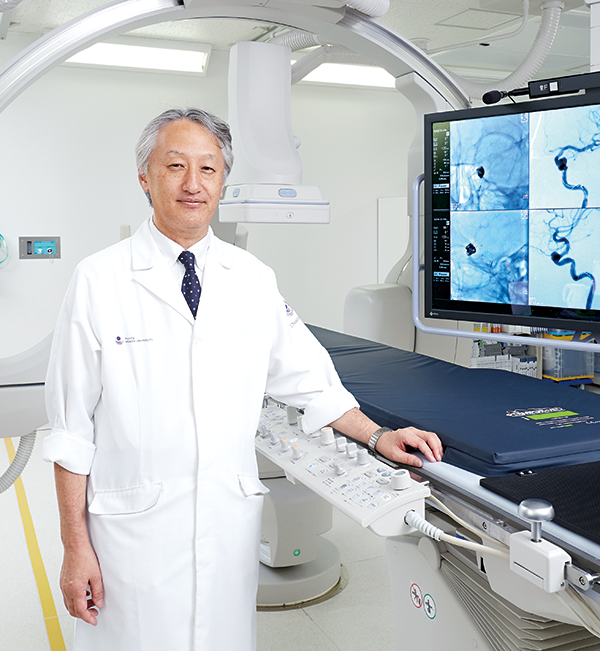 Prof. Ichiro Nakahara / Department of Comprehensive Strokology / Fujita Health University Hospital
It’s no exaggeration when I say the Alphenix Hi-Def Detector and Alphenix Workstation have entirely changed the clinical practice for me and my team – not to mention the thousands of patients we treat every year.
Until recently, stroke treatment at our hospital had always been delivered in a nonintegrated silo: only the neurosurgical team was managing surgical treatment, and the department of neurology was managing all things medical. However, with neuroendovascular therapy growing and awareness increasing, the need for a more integrated approach was becoming evident. It was clear that in order to strategize the future of our practice, we needed technology that could bridge the divide between surgery and medicine, as well as provide patients with the best-in-class neuroendovascular treatment they need.
We currently have five hybrid operating rooms, each equipped with an angiography system from various vendors. Of all the existing systems, I mainly prefer to use the Alphenix Biplane system manufactured by Canon Medical Systems Corporation, which was installed at our hospital for imaging the head and neck.
Until recently, stroke treatment at our hospital had always been delivered in a nonintegrated silo: only the neurosurgical team was managing surgical treatment, and the department of neurology was managing all things medical. However, with neuroendovascular therapy growing and awareness increasing, the need for a more integrated approach was becoming evident. It was clear that in order to strategize the future of our practice, we needed technology that could bridge the divide between surgery and medicine, as well as provide patients with the best-in-class neuroendovascular treatment they need.
We currently have five hybrid operating rooms, each equipped with an angiography system from various vendors. Of all the existing systems, I mainly prefer to use the Alphenix Biplane system manufactured by Canon Medical Systems Corporation, which was installed at our hospital for imaging the head and neck.
A strategic solution for the long term
In March 2019, we had decided to install the Alphenix Hi-Def Detector and Alphenix Workstation.
As a neurovascular surgeon, it is my job to perform both neurosurgery and neuroendovascular therapy, and I can tell you from experience, the new Alphenix platform solution have changed the way we practice intervention for us. Here’s why:
Seeing device details can be incredibly effective.
The Alphenix Hi-Def Detector has achieved the world's smallest pixel size (at 76 µm) for a detector used in any angiography system. To put this in context, this is more than half the pixel size of any other detector in the market, allowing the system to provide high-definition images in both fluoroscopy and radiography. Now, a field size as small as 1.5 inches (3.89 cm × 3.89 cm), which is the minimum FOV of the Alphenix Hi-Def Detector, can be enlarged and displayed as extremely sharp images on the large monitor.
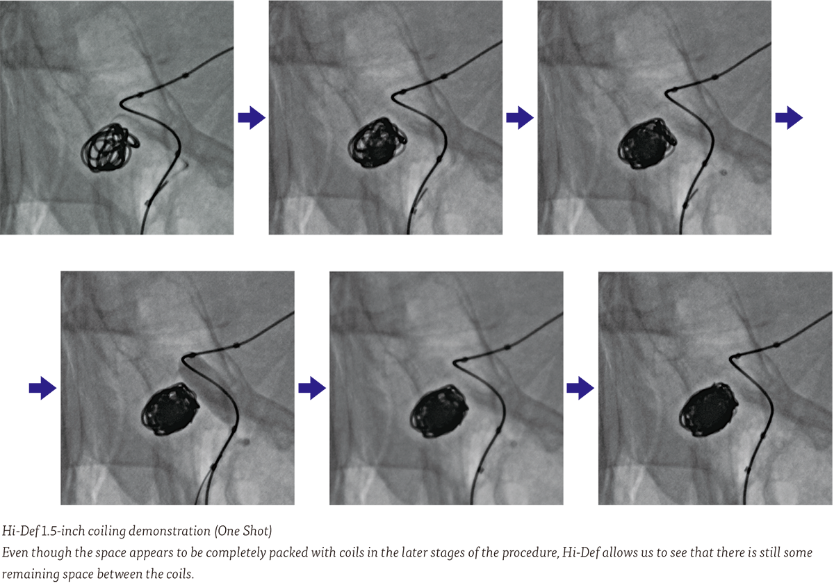
Enhances our surgical case experience
I first used a prototype of the Alphenix Hi-Def Detector about two years ago and was amazed at how clearly it could depict small microcatheters. I had the same feeling when I used the system installed at our hospital. The position of a microcatheter introduced into an aneurysm can be precisely determined, and the coils placed in the aneurysm can be distinctly visualized. Indeed, the ability to view the placement of the framing coil in the aneurysm is far superior to anything we had experienced in the past. In the course of our practice, we often employ flow-diverter stents for large/giant aneurysms, and the detector allows even the finest struts of such stents to be visibly depicted.
I believe that the main advantage of the Alphenix Hi-Def Detector is its high definition zoom capability, which makes it possible to observe extremely small structures in enlarged views (down to 76 µm pixels) on the large monitors and ensures extremely fine control by the surgeon for increased precision.
Assists us with treating cerebral aneurysms
By using the Alphenix Hi-Def Detector, it makes it possible to increase the packing density in coil embolization procedures for aneurysms. Even when surgeons feel that much of the space within an aneurysm has been packed with coils, viewing an enlarged Hi-Def detailed image of the aneurysm often reveals remaining space for the placement of additional coils.
It is my opinion that the coil packing ratio of aneurysms could be increased from the usual 30% to around 40%, or even 50%, by employing this simple technique. This, of course, requires further study for verification, but I am planning to conduct research on this matter in the future.
During postoperative follow-up, the use of the Alphenix Hi-Def Detector can increase accuracy in evaluating recanalization. And in the placement of flow-diverter stents, the images acquired using the Alphenix Hi-Def Detector are totally different visual experience from those acquired using a conventional detector. When the Alphenix Hi-Def Detector is used, it is possible to place the stent at the optimal position in closer contact with the vessel wall, and even small perforating vessels to be conserved can be depicted, allowing us to determine the optimal position for placement of the flow-diverter stent.
Outside Japan, a new endovascular flow disruptor device, known as the WEB (Woven Endo Bridge) by Microvention, for reducing blood flow within an aneurysm has been introduced in clinical practice, and it is anticipated that this device will also be introduced in clinical practice in Japan within the next few years. Alphenix’s Hi-Def imaging would also be useful for further enhancing such novel treatment methods, and the Alphenix Hi-Def Detector is expected to play a significant role in this field.
Its potential future for neuroendovascular therapy.
The ability of the Alphenix Hi-Def Detector to provide Hi-Def images may expand the application of neuroendovascular therapy to treat finer vessels in the future. In addition to the treatment of the cerebral arteries as discussed above, the detector may also help physicians accurately determine the extent of shunt flow in a variety of conditions, such as cerebral arteriovenous malformations and dural arteriovenous fistulas (d-AVF).
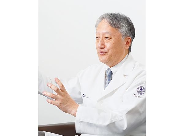
“Only after experiencing cases in Hi-Def, you truly appreciate the detailed visual and clinical advantages it provide. Now it’s difficult to go back using a conventional detector again.”
Professor Ichiro Nakahara
Department of Comprehensive Strokology
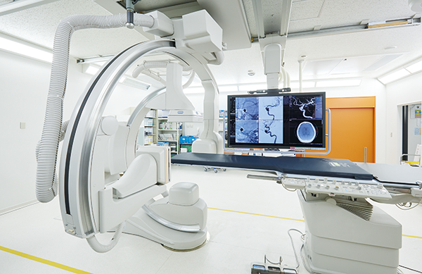
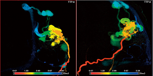 Diagnosis of AVM using PI
• The working angles of the biplane system can be automatically set in advance by simply pressing a button on the workstation, or new tablet.
• The Parametric Imaging (PI) function uses the time-density curve for each pixel acquired by pre-and post-digital subtraction angiography (DSA) to display the changes in pixel values in different colors according to characterize the contrast media dynamics and to allow easier visual flow evaluation in a single composite image.
• The Parametric Imaging (PI) function uses the time-density curve for each pixel acquired by pre-and post-digital subtraction angiography (DSA) to display the changes in pixel values in different colors according to characterize the contrast media dynamics and to allow easier visual flow evaluation in a single composite image.
• Color-Coded Circulation (CCC) is a function that simultaneously shows both the anatomical course of blood vessels and blood flow by displaying color information as a dynamic image based on the contrast medium arrival time.
I believe that the effectiveness of neuroendovascular therapy could be evaluated by using both PI and CCC in preoperative and postoperative imaging.
• In Cerebral Aneurysm Analysis (CAA), aneurysms are semi-automatically detected in 3D images acquired using Alpha CT (cone-beam CT imaging). When the neck of an aneurysm is selected, the parent blood vessel is segmented and the measurement results are displayed. The size and volume of the aneurysm are measured automatically, and the results can be used to evaluate the embolization ratio.
The Workstation and Hi-Def Detector integrate beautifully
The images acquired using Alpha CT (CBCT) with the Alphenix detector are significantly superior to those acquired using other conventional detectors, and I look forward to additional progress in the future. It is my firm hope that the Alphenix Hi-Def Detector will undergo further development to achieve high-definition Alpha CT (CBCT) using the high-definition detector technology. Such high-definition 3D images would lead to more accurate diagnosis and treatment for clinicians.
Concerning workstation functions, such as CFD (computational flow dynamics analysis) and 4D-Perfusion (flow dynamics analysis on the temporal axis), these could be further improved. Ultimately, it would be advantageous if the entire course of stroke care, including all the steps in evaluating and determining infarction, neuroendvascular treatment, and post treatment follow-up could be conducted with solely the angiography system used for stroke patients admitted in emergency cases.
Looking to the future
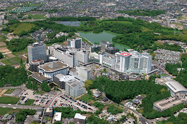 Fujita Health University Hospital
As a user of an Alphenix Biplane system, I feel that the angiography systems manufactured by Canon Medical, including those with the Alphenix Hi-Def Detector and the Alphenix Workstation, have undergone dramatic improvement recently.
Although I have experience using angiography systems from other manufacturers, I currently feel very comfortable and relaxed using our Alphenix system.
It is reasonable to say that the Alphenix Hi-Def Detector, which provides very sharp images even at a minimum FOV of 1.5 inches, is an essential feature for physicians who perform neuroendovascular procedures, as even a single millimeter matters in the treatment of cerebral aneurysms. It is my belief that the Alphenix Hi-Def Detector and the Alphenix Workstation will play leading roles in the further development of neuroendovascular therapy in the future.//
Although I have experience using angiography systems from other manufacturers, I currently feel very comfortable and relaxed using our Alphenix system.
It is reasonable to say that the Alphenix Hi-Def Detector, which provides very sharp images even at a minimum FOV of 1.5 inches, is an essential feature for physicians who perform neuroendovascular procedures, as even a single millimeter matters in the treatment of cerebral aneurysms. It is my belief that the Alphenix Hi-Def Detector and the Alphenix Workstation will play leading roles in the further development of neuroendovascular therapy in the future.//
Social Media


