

Supporting Advances in Gynecologic Oncology
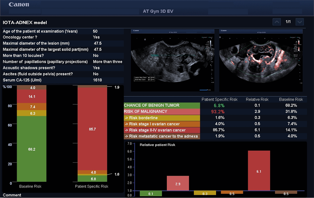
IOTA-ADNEX model in case of advanced stage Ovarian Cancer.
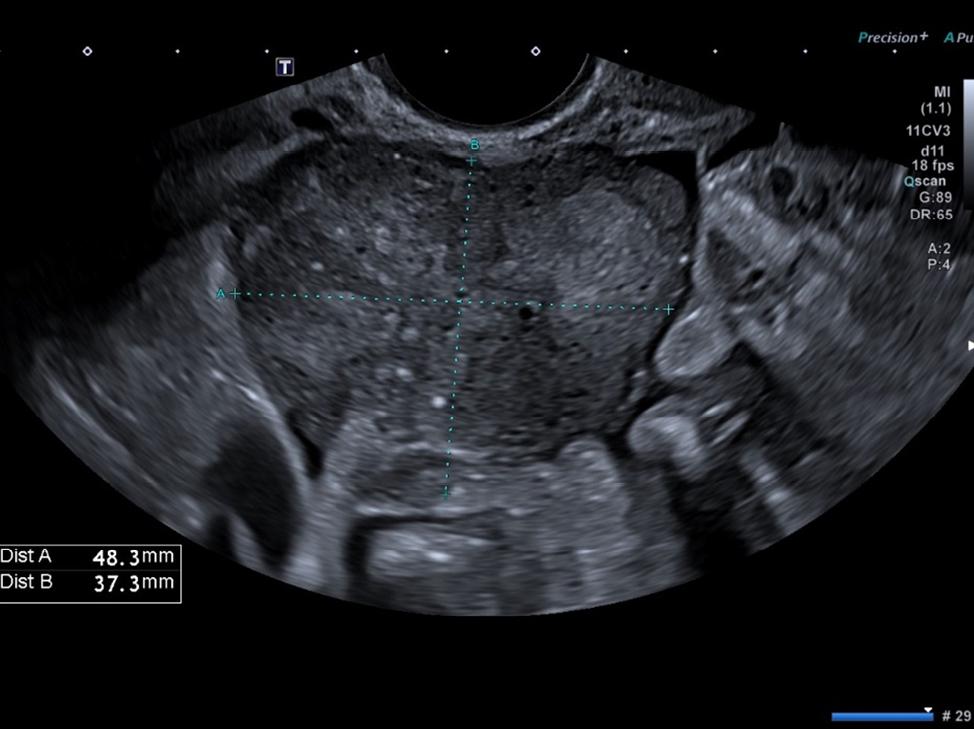
Ovarian Cancer.
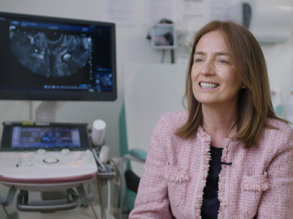
“In addition, through working as part of the gynecology team, I realized the great value of ultrasound in the daily clinical management of our patients. In particular, during follow-up, when we can use ultrasound as a bedside tool. For example, if we have a suspicion of a recurrence, we can check it immediately to understand the nature of one or more lesions and assess the possibility to surgically remove the lesion. In addition we can obtain a biopsy of that lesion to follow up the situation during treatment,” she continued.
“I wanted to share my knowledge with my colleagues, with residents and with students, which is why I created the Center for Ultrasound in Gynecological Oncology “Class Ultrasound” at the Agostino Gemelli University Hospital Foundation in Rome, Italy. Since its inception six years ago, around 2,500 colleagues have attended courses there.” The Center has trained Italian and European physicians and health professionals, provides patients with the highest professional service and plays a key role in multi-center scientific studies.
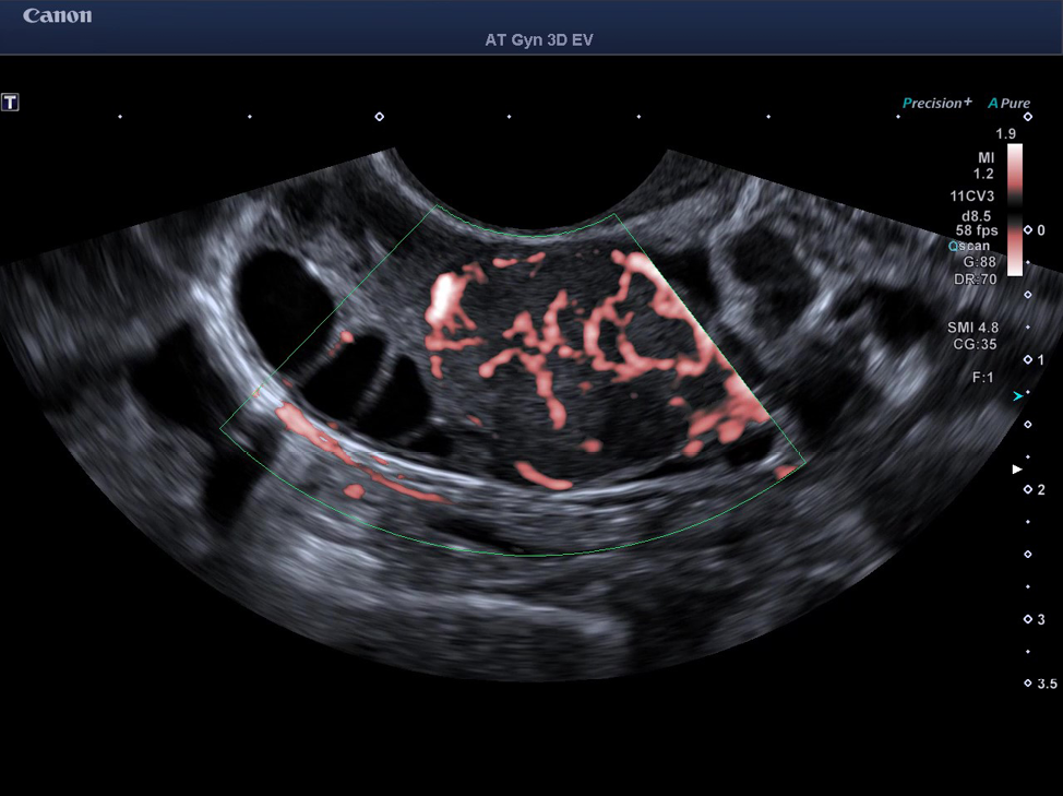
Ovarian Cancer.
“The most important requirements relating to an ultrasound system that I am always looking for are first - resolution, so high-quality grayscale images. Then, intuitive software, that allows the examiners to use the equipment at its best. In other words, easy-to-manage tools that are not very complicated to use. Then, high-quality vascularization assessment,” she continued. “When I discovered Aplio ultrasound machines, I was fascinated by its very high image quality with crystal-clear images and enhanced penetration. Then I found the equipment very good, able to deliver superb images for a wide range of clinical applications. Moreover, regarding the vascularization, it was amazing to find that the machine had the possibility to detect very, very small vessels, which is crucial for us to discriminate papillary projections or amorphous material, for instance, within an ovarian cyst. I have to say that also the Doppler Luminance is fantastic, because it gives the impression of 3D rendering images.”
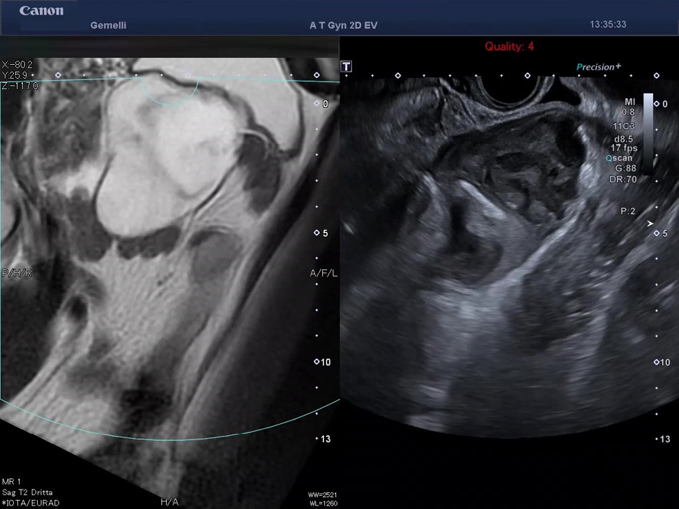
Smart Fusion Image of Low-Grade Appendiceal Mucinous Neoplasm.
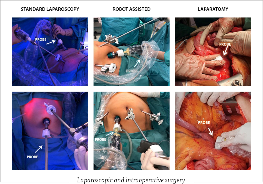
“Secondly, the challenge is to have a bedside tool to get a quick assessment to solve some clinical problems, to check for pleural effusion and presence of ascites or do simple procedures, such as paracentesis, drainage of fluid or place intraperitoneal catheters. And thirdly, we discovered that ultrasound can play an important role in the surgical field. As our patients very often had been already treated with radiotherapy, and/or previous surgical procedures, sometimes, it's very challenging to detect a small recurrent lesion located in the lower part of the pelvis, for example. With the transvaginal transducer, we can guide the surgeon to reach that small area. Besides that, Canon Medical provided us with a laparoscopic ultrasound transducer, and this is fantastic, because for some patients with borderline ovarian tumors, for instance, we are able to provide ‘fertility-saving’ surgery. We can select the area and localize the small lesion in order to spare the largest amount of normal tissue.”
New research possibilities
The systems have also enabled new possibilities in research. “As far as innovation is concerned, I was very interested in the possibility to explore Smart Fusion that fuses Ultrasound with MR or CT images. So, we decided to run a prospective study on patients with cervical cancer and a study in patients with advanced ovarian cancer. We have already published the results on the first study - cervical cancer. We have been analyzing data of a large series of patients with ovarian cancer,” said Prof. Testa. “At the moment, I can say that I'm very happy about this experience and, in my opinion, Smart Fusion can play a prominent role in education. To have the possibility to share the experience, being the radiologist and gynecologist together the same room - It's very important for the improvement of the experience of both disciplines.”
Working on the future
Canon Medical continuously develops its ultrasound systems to provide even better image quality and workflow that enable clinicians to advance medicine. One important aspect is integrating Artificial Intelligence (AI) enabled technologies into the ultrasound systems in the future.
“I look confidently towards the future,” added Prof. Testa. “I really hope that ultrasound equipment will provide us with Artificial Intelligence analysis. With this, we will be able to store a lot of images using a simple method plus define and recognize more of what we cannot do with our eyes and brains.” //
Publications:
1 Intraoperative ultrasound diagnosis of metastatic lymph node in serous borderline ovarian tumor De Blasis I, Tortorella L, Macchi C, Arciuolo D, Scambia G, Testa AC (UOG 2019). https://obgyn.onlinelibrary.wiley.com/doi/10.1002/uog.20234
2 Ultrasound Technologies in the diagnosis and treatment of ovarian cancer Antonia Testa, Canon Medical webinar at ISUOG World Congress 2020. https://www.youtube.com/watch?v=-4H9Prl37UM
3 Fusion imaging of ultrasound and MRI in the assessment of locally advanced cervical cancer: a prospective study Moro F, Gui B, Arciuolo D, Bertoldo V, Borzi R, Romeo P, Petta F, Cambi F, Pasciuto T, Zannoni GF, Valentini V, Manfredi R, Scambia G, Testa AC Italy (IJGC 2019). https://ijgc.bmj.com/content/30/4/456
Social Media


