

Deep Learning Reconstruction in Clinical MRI:
a new realm combining ultra-high resolution and increased productivity

Garry E. Gold, M.D., Ph.D.
Clinical radiologist and researcher
Past president of the International Society for Magnetic Resonance in Medicine (ISMRM) and
the Society of Computed Body Tomography and Magnetic Resonance (SCBT/MR)
Introduction
One of the main challenges in MRI is finding the optimal balance of Signal to Noise Ratio (SNR) and Image Resolution such that detailed structures can be clearly visualized. While nearly every radiologist dreams of ultrahigh resolution, the reality remains that when spatial resolution is increased, the available signal per voxel decreases, leading to lower SNR. Image noise can then hinder visualization of critical features and introduce uncertainty and frustration into the diagnostic process. When noise becomes overly apparent, technologists have no choice but to manipulate parameters, commonly leading to both protocol inconsistency and prolongation of scan times. These undesired consequences negatively affect patient care and decrease productivity in the radiology department.
New developments in Artificial Intelligence (AI) have led to advances in Deep Learning Reconstruction (DLR). Canon Medical’s Advanced intelligent Clear-IQ Engine (AiCE) is a deep-learning based solution trained with specific datasets to learn to detect noise and remove it from clinical images, permitting spatial resolution to be increased or acquisition time to be reduced.
In accordance with Canon Medical’s goal of improving patient care, the initial approach to the adoption of AI in MRI was clear: reduce examination times, enhance patient comfort, expand diagnostic capabilities, and enrich every radiologist’s confidence. These outcomes have never been mutually achievable before – we are truly witnessing a new era in Magnetic Resonance.
Advanced intelligent Clear-IQ Engine (AiCE):Redefining MRI relationships
In MRI, image resolution is defined by pixel size and slice thickness. The depiction of visually distinct anatomical structures is dependent upon those spatial factors, as well as noise and apparent image contrast. The higher the spatial resolution of an image (small pixels and thin slices) the better the detail, but the lower the SNR, since the smaller pixels and thin slices contain less signal from protons.
Noise, which manifests itself as graininess in the image, is random and always present in nature. When excessive, noise can diminish the human eye’s ability to detect edge detail and dull contrast between tissue types. A common way to increase SNR while maintaining high resolution is to acquire several signal measurements (“Number of Acquisitions” or “NAQ”) and average them. Because of its random fluctuations, averaged noise in one location does not increase as fast as signal values. Thus, the process of acquiring multiple averages is a robust way to improve image quality at a cost of increased scan time.
Inspired by the architecture of the human brain, AiCE DLR uses a Deep Convolutional Neural Network (DCNN) capable of both learning and performing complex tasks when working with image data1.
During the DCNN’s training process, computational models were created by analyzing a large dataset of examinations acquired on several subjects’ brains and knees. These models included the analysis of a wide array of contrast weightings (T1, T2, FLAIR, PD), thus ensuring that AiCE DLR would be efficient and robust across multiple clinical contexts.
To perform this training, system input of ideal SNR high resolution datasets with 10 NAQ were defined as “ground truth” and then mixed with Gaussian noise. This data was then processed by the neural network, which was built in such a way as to automatically define the noise level and to propose a denoising solution1 Figure 1.
The resulting low noise images were then compared to native images. If there were different, the network learned, adjusted the algorithm weights, and the denoising process repeated. At the end of the training procedure, the denoised images and the ground truth images were considered similar, algorithm weights were saved and the trained database completed.
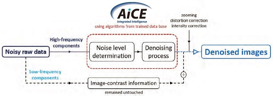
While the training procedure itself is time-consuming, Canon Medical’s clinical application of DLR algorithm is fast and efficient since the DCNN can simply apply the pre-existing knowledge of high-SNR, high-quality data analyzed during its learning period. Denoising occurs in the spectral domain where only the high frequency content is passed through the network. Adaptive activations automatically adjust to the input image noise level, producing high-quality images across all sequences without requiring any user interaction, thus efficiently optimizing the removal of noise while preserving key MR signals Figure 2. After denoising, the low-frequency components containing the image-contrast information have remain untouched and are combined with the optimized results in order to maintain native contrasts. Interpolation and distortion/intensity corrections are finally applied following the DLR process, thus completing image reconstruction.
» AiCE changes the way we think about MRI.«
Garry E. Gold, M.D., Ph.D.
Figure 1 Training procedure leading to the creation of a robust database.
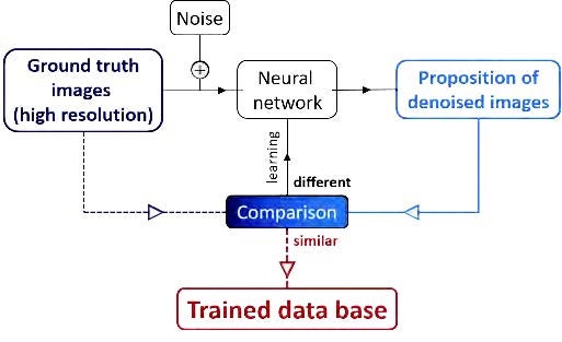
Figure 2 Denoising process in the clinical context. Noisy raw data is quickly and automatically evaluated using the trained algorithm, resulting in denoised images.
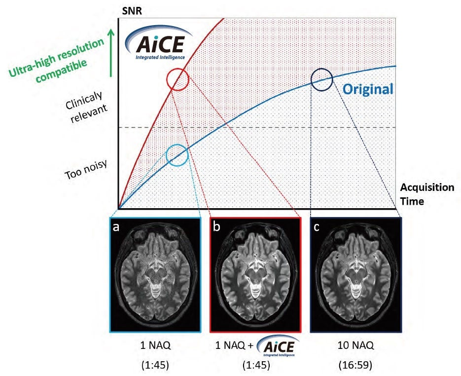
Since the introduction of AiCE DLR, typical relationships of SNR and scan time have been redefined. Figure 3 illustrates a comparison between traditional images and those with AiCE DLR. While non-averaged T2 - weighted images (a) appear too noisy to be clinically relevant, the same dataset processed with AiCE DLR (b) exhibits comparable quality to a ten-times averaged acquisition (c), without requiring the excessive scan time traditionally inherent to such a scenario. These striking results change the way we think about MR imaging, revealing new pathways for providing ultra-high resolution in the clinical setting without sacrificing SNR or workflow. Preliminary studies have tested the potential of the technique when applied to several clinical contexts2-4.
» I’m impressed by the ease-of-use, how it maintains image contrast and structural detail, while at the same time eliminating noise.«
Garry E. Gold, M.D., Ph.D.
Figure 3 Relationships between SNR, acquisition time, standard processing, and AiCE DLR are illustrated with routine T2 - weighted axial brain images. Single average original images (a). Same images post-processed with AiCE DLR (b) demonstrating quality strikingly similar to that of an acquisition using 10 NAQ (c), at a fraction of the scan time.
Achieving Ultra- High Resolution with average scan times at 3T
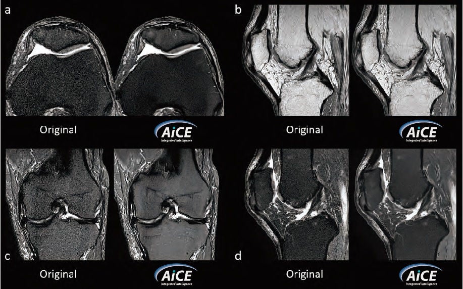
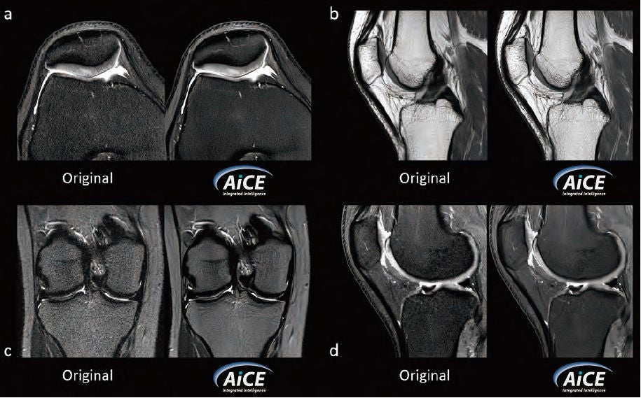
AiCE DLR denoising tool benefits clinical practice since it allows scanning at a newly defined ultra-high resolution while maintaining routine scan times. By using a protocol built specifically for DLR, with interpolated pixel sizes of 100-200 μm, slice thicknesses of 1.5 mm and scan time of less than 6 minutes, it is possible to compare the results of images processed using AiCE DLR and those with standard processing Figure 4. Images on the left are considered to be of non-diagnostic quality, due to the amount of noise present, but the number of NAQ necessary to improve them would have increased scan time to a point no longer considered clinically feasible. AiCE DLR images on the right demonstrate a remarkable reduction of noise, while contrast and fine structural detail can be easily characterized.
Figure 4
Original (left) and AiCE DLR (right) MRI of a knee with ultra-high resolution and average scan times.
(a) Ax PD FS ; Interpolated resolution = 100×100 μm² ; Slice thickness = 1.5 mm ; Acquisition time = 4:45
(b) Sag PD ; Interpolated resolution = 100×100 μm² ; Slice thickness = 1.5 mm ; Acquisition time = 4:58
(c) Cor STIR ; Interpolated resolution = 200×200 μm² ; Slice thickness = 1.5 mm ; Acquisition time = 5:34
(d) Sag PD FS ; Interpolated resolution = 100×100 μm² ; Slice thickness = 1.5 mm ; Acquisition time = 4:53
Achieving Ultra-High Resolution with short scan times in 3T
Another approach to the use of AiCE DLR is to combine decreased scan times and very high spatial resolution. In Figure 5, pixel size was reduced to 200 μm, slice thickness of 3 mm, and an acquisition time of two minutes or less, allowing a routine knee to be completed in under 8 minutes. When compared with the average 20-30 minutes-long, routine knee scan, the AiCE DLR knee approach provides similar image quality, at an even higher resolution in a fraction of the time. These types of protocols can be used not only for increased productivity in the radiology department, but also for providing improved care to a specific patient segment such as children and the elderly. For these patients, faster scan times might be the only way that diagnostic results can be obtained. By allowing higher spatial resolution at shorter scan times, AiCE DLR is able to change the MRI environment for doctors and patients alike, since better diagnosis, accessibility and productivity can only translate to improved patient care.
Figure 5
Original (left) and AiCE DLR (right) MRI of a knee with ultra-high resolution and short scan times.
(a) Ax PD FS ; Interpolated resolution = 200×200 μm² ; Slice thickness = 3 mm ; Acquisition time = 1:55
(b) Sag PD ; Interpolated resolution = 200×200 μm² ; Slice thickness = 3 mm ; Acquisition time = 2:00
(c) Cor STIR ; Interpolated resolution = 200×200 μm² ; Slice thickness = 3 mm ; Acquisition time = 1:58
(d) Sag PD FS ; Interpolated resolution = 200×200 μm² ; Slice thickness = 3 mm ; Acquisition time = 2:00
Disclaimer: The clinical results, performance and views described in this document are the experience of the health care providers. Results may vary due to clinical setting, patient presentation and other factors. Many factors could cause the actual results and performance of Canon Medical’s product to be materially different from any of the aforementioned.
Conclusions
The use of AI in medical imaging continues to be a hot topic in today’s clinical radiology and research communities. However, AI has come a long way since its introduction and it is imminent that, in addition to serving as a diagnostic aid, it will be eventually implemented in all areas of our industry (i.e.: manufacturing, patient safety, registration, and post-processing). With the introduction of AiCE DLR in MRI, Canon Medical has taken the first step towards solving one of the modality’s biggest challenges, with evident benefits to patient care.
» With AiCE, we can easily address the two most pressing clinical needs of our time: superb image quality with ultra-high matrices acquired within routine scan times or abbreviated scan times with traditional resolution. These combinations, which could not previously be achieved, now allow radiologists to better tailor scans to patient needs, improving both patient care and departmental productivity.«
Garry E. Gold, M.D., Ph.D.
References:
- Masafumi Kidoh, Kensuke Shinoda, Mika Kitajima, Kenzo Isogawa, Masahito Nambu, Hiroyuki Uetani, Kosuke Morita, Takeshi Nakaura, Machiko Tateishi, Yuichi Yamashita, Yasuyuki Yamashita “Deep Learning Based Noise Reduction for Brain MR Imaging: Tests on Phantoms and Healthy Volunteers.” Magnetic Resonance in Medical Sciences, 2019. https://doi.org/10.2463/ mrms.mp.2019-0018.
- Mitsue Miyazaki, Masaaki Umeda, Shinichi Kitane, Cheng Ouyang, Sheronda Statum, Won C Bae, and Christine Chung “Scan Time Reduction in High Resolution Knee Imaging using Compressed Sensing and Denoising Deep Learning Reconstruction.” ISMRM Congr. 2019.
- Ryuichi Mori, Hideki Ota, Atsuro Masuda, Tomoyoshi Kimura, Tatsuo Nagasaka, Takashi Nishina, Sho Tanaka, Yoshimori Kassai, and Kei Takase “Ultrashort TE Time-Spatial Labeling Inversion Pulse MR Angiography Denoised with Deep Learning Reconstruction for Abdominal Visceral Arteries: A Feasibility Study.” ISMRM Congr. 2019.
- Sanae Takahashi, Haruhiko Machida, Toshiya Kariyasu, Rieko Niitsu, Isao Miyazaki, Tatsuya Yoshioka, Keita Fukushima, Saori Yuda, Yuta Shimizu, Takayuki Yonaha, Akihito Nakanishi, Hiroshi Kusahara, Youhei Matsuoka, Miho Kitamura, Takao Yamamoto, and Kenichi Yokoyama “Improved vessel delineation in whole-heart coronary MRA with sub-millimeter isotropic resolution using deep learning reconstruction compared with routine whole-heart coronary MRA.” ISMRM Congr. 2019.
Clinical and Scientific Canon Medical support


Social Media


