

Adjusting Contrast Media Dose to Compensate for Changes in kVp During Portal Phase Liver CT

Appropriate contrast enhancement of organs in CT is key to promoting accurate diagnoses. With abdominal imaging it is particularly important to ensure the liver is appropriately enhanced as cancers may metastasise within it. As the vast majority of the hepatic blood supply comes from the portal vein, the portovenous (parenchymal) phase is often the most diagnostically useful due to the tissue being at its most homogenous, making anomalies easier to distinguish.
Many factors affect contrast media enhancement. Much has been written on individual contrast dosing, e.g. weight-based and also accurate scan delay times, but an area often overlooked is the effect from the choice of kVp used. With the advent of more efficient detector technology alongside iterative reconstruction techniques, CT exposure levels have decreased, so this has led to the opportunity to employ lower kVps. Altering the kVp with a corresponding amendment of the modulated tube current (mA) can achieve a constant image quality in terms of signal to noise ratio, but there are considerations that need to be understood before this is done:
a) The patient dose may be affected despite the overall image quality being maintained, i.e. lowering the kVp with a corresponding (possibly automated) raise in mA to maintain image quality, may actually deliver a higher patientdose.
b) All X-ray tubes have a maximum mA achievable. Lowering the kVp may result in the mA ‘topping out’, i.e. to maintain image quality the scanner would need to deliver more mA than the tube can achieve. Continuing with this scan may therefore result in an under exposure and poor signal to noise ratio. This is more often a consideration before lowering the kVp for larger patients.
c) The lower the kVp the stronger the effect will be from IV contrast media. Bae 20101 discusses that using lower tube voltages will result in higher CT attenuation because the X-ray output energy is closer to the iodine k edge of 33 keV and that iodine concentration of 1 mg of iodine per milliliter corresponds to contrast enhancement of approximately 30 Hounsfield Units for 100 kVp but 40 Hounsfield Units for 80 kV. This confirms that we should take kVp into consideration what deciding upon a suitable contrast dose to administer.
Generally only a small number of kVp settings are available. Using the Canon CT system, the Aquilion ONE / GENESIS Edition, for example there are four – 80, 100, 120 and 135. Routinely, we seek to use 100 kVp for adult abdominal imaging. For larger patients however, we may need to switch up to 120 kVp to ensure we deliver an adequate X-ray exposure to ensure image quality. When doing this we instruct the radiographers to make an additional adjustment to the contrast media delivered to try to maintain adequate liver enhancement. Conversely, for patients with poor renal function, we sometimes switch down to 80 kVp which enables us to reduce the contrast dose delivered whilst still maintaining adequate enhancement, and thereby help protect the kidneys.
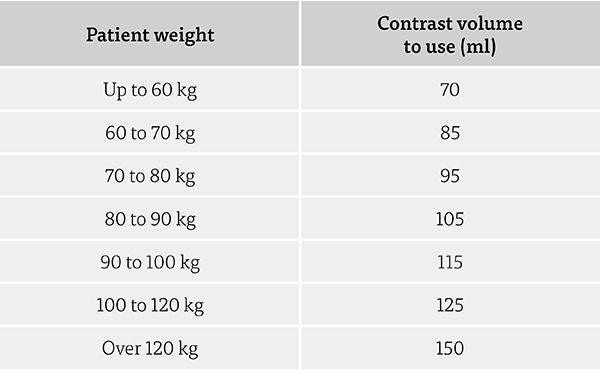
Figure 1.
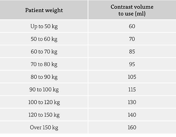
Figure 2.
Study
The optimum portovenous liver enhancement had already been decided in a previous study to be 100-125 HU1. In bottled concentration, contrast media enhances considerably more than this of course, but during the portal phase it will have been significantly diluted with blood and shared throughout the body such that only a proportion will be within liver. So a phantom was required to simulate a liver loaded with optimal portovenous amount of contrast media. According to Andersen et al 20003, the average liver volume is around 1.3 to 2 litres, and so 1.5 litres of water were poured into a radiolucent vessel (sharps bin – Figure 3) and then small amounts of 350 strength contrast media were added and it was repeatedly scanned, firstly at 80kVp until an optimal 115 Hounsfield Units (HU) were measured. The solution was then rescanned at 100, 120 and 135 kVp and each enhancement recorded (Figure 4).
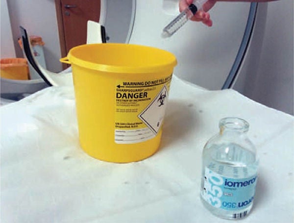 Figure 3.
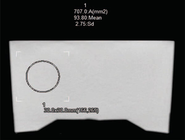 Figure 4.
Next, more contrast media was added until 115 HU was achieved at 100 kVp. The solution was then scanned at 80, 120 and 135 kVp and the enhancements again recorded. This was repeated twice more by achieving 115 HU enhancement at 120 and 135 kVp. Whilst this is admittedly a crude phantom, it was felt that it represented a similar sized volume of fluid to a liver, infused with an amount of contrast media to offer desirable in-vivo portal phase enhancement, i.e. contained a similar amount of iodine per volume.
Results
A target of 115 HU was chosen to be considered mid-range, and therefore optimum portovenous enhancement.
At 80 kVp it was found that it required 11 mls of 350 mgI/ml contrast media to be added to the 1500ml water to result in a CT enhancement of 115 HU. This mixture was then scanned at 100, 120 and 135 kVp and the HU measured and noted.
An additional 4mls of 350 mgI/ml, i.e. a total of 15 mls was required to reach 115 HU at 100 kVp. So as expected a stronger solution was required to maintain enhancement at 100 kVp compared with 80 kVp. This new mixture was then scanned at 80, 120 and 135 kVp and the HU measured and noted.
This process was repeated at 120 kVp which required a total of 19 mls of contrast media, and then at 135 kVp which required it to be increased to 22 mls of contrast media.
The results are in tabulated in Figure 5.
 Figure 5.
So it can be seen that the solution strengths required to maintain 115 HU enhancement at each kVp were:
- 80 kVp – 350 x 11 mgI, or 3.85 g iodine in 1511 ml fluid, or 1 g in every 392 ml of solution
- 100 kVp – 350 x 15 mgI, or 5.25 g iodine in 1515 ml fluid, or 1 g in every 288 ml solution
- 120 kVp – 350 x 19 mgI, or 6.65 g iodine in 1519 ml fluid, or 1 g in every 228 ml solution
- 135 kVp – 350 x 22 mgI, or 7.7 g iodine in 1522 ml fluid, or 1 g in every 198 ml solution
So, at 135 kVp TWICE the iodine concentration (contrast dose) is needed to maintain the same enhancement as at 80 kVp.
How would we put this into practice to ascertain what changes we should actually make when needing to (or choosing to) raise or lower kVp?
Example 1
Assume we are to scan a large patient who requires a change from 100 kVp to 120 kVp. How much contrast media increase should be to be injected to result in the appropriate amount of contrast reaching the liver, and maintain the desired portovenous phase enhancement of 115 HU?
Let us assume the patient is 85 kg. Using our current weight-based look-up table we would inject 105 mls at 100 kV, with the aim to enhance the liver to 115 HU. From the results we can now assume that we would be loading the liver with 5.25g (or more if a larger liver) of contrast media, equating to 1mg for every 288mls of fluid held in the liver.
So lets us assume that our scanner mA modulation has told us that we are going to need to go up to 120 kVp to maintain image quality. We need to therefore load the liver with around 6.65 or more mg Iodine (depending on liver volume), i.e. 1 mg for every 228 mls of solution in the liver.
Therefore this suggests that we should inject proportionally more contrast media, i.e. 288 / 228 x 105mls = 133 mls of 350 strength contrast media.
Using our standard rules we would have only increased by one weight category for a kVp increase, i.e. we would have given 115 mls, but this shows that in fact we may achieve better imaging by increasing two weight groups to give 130 mls.
This would be a contrast dose increase of around 25%.
Example 2
We are about to scan a patient at 100 kVp, but have established a poor renal function, so decide to drop to 80 kVp such that we can give less contrast media whilst maintaining enhancement. How much can we drop by? Using our current system we would drop by one group.
Let’s assume the patient is 65 kg. She would usually receive a weight-based dose of 85 mls to attempt to enhance the liver to around 115 HU. This equates to a dose of 1mg iodine for every 288 mls fluid, but at 80 kVp the results suggest that she would need only 1 mg iodine in 392 mls fluid. Therefore, this advocates we should be able to drop by 288/392 x 85 mls = 62 mls and maintain enhancement. Using our current rule of dropping one group, we would have only reduced to 75 mls, but in actual fact we could do much better by dropping two groups to give 60 mls. This would be a contrast reduction of around 30%.
So in general, we can infer some multiplication factors for kVp changing that should maintain the same portal phase enhancement as follows:
- Change from 80 > 100 kVp, increase volume by x 1.36
- Change from 100 > 120 kVp, increase volume by x 1.26
- Change from 120 > 135 kVp, increase volume by x 1.15
- Change from 135 > 120 kVp, decrease volume by 0.87
- Change from 120 > 100 kVp, decrease volume by 0.79
- Change from 100 > 80 kVp, decrease volume by x 0.73
As our default kVp is 100, the most useful two changes for us to consider are raising from 100 to 120 kVp for larger patients (we practically never need to go as high as 135 kVp), and, lowering from 100 to 80 kVp for patients with poor renal functions (though very low kVp is often only a realistic option for smaller patients where an adequate overall exposure can still be attained).
This table in Figure 6 shows that when looking to lower the kVp (green arrows) for lighter patients, for those under 60 Kg a change of one weight category (as currently done) is enough. However for middle weight patients (60 to 80 Kg) a change of two weight groups was likely to give a perfectly adequate enhancement.
When needing to raise the kVp for heavier patients (over 90 Kg) adjusting the amount delivered by two weight categories rather than one was also likely to offer better enhancement.
Practically, I therefore decided to implement a new rule to change by two weight categories instead of our existing rule of one, keeping the minimum adult delivery to 60 mls, and maximum as 160 mls.
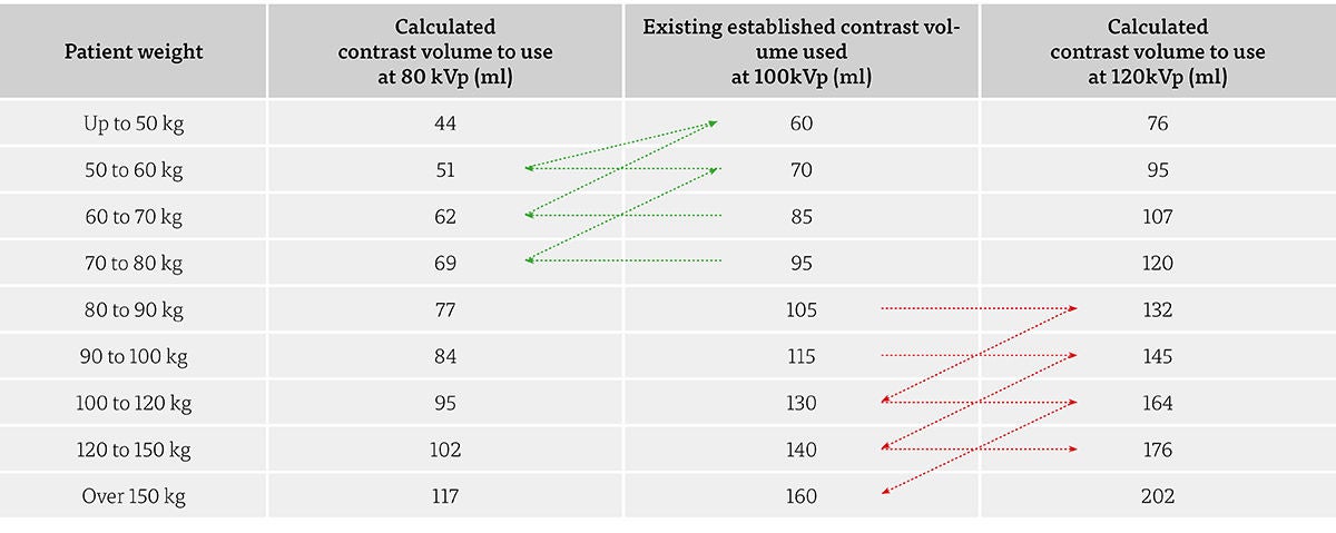 Figure 6.
Summary
Altering the kVp used for portovenous CT imaging has a significant effect on the contrast enhancement achieved. Adjustments in contrast dosing should be made to compensate where a change in kVp is undertaken – increase contrast dose for higher kVp, decrease contrast dose for lower kVp. The amount adjusted will likely vary for differing scanners as beam filtration is not the same for all vendors or scanner models. In this study, for our system, a change up from 100 kVp to 120 kVp required 25% more contrast to maintain the same enhancement. Similarly a change down from 100 kVp to 80 kVp allowed a 30% drop in contrast dose to maintain enhancement. The adjustment needed seems to be larger than we previously had been making. We have subsequently changed our practice in line with these findings.
Interestingly, a hypothetical adjustment (as we would never need to do this) from 80 kVp to 135 kVp would require somewhere in the order of a doubling of contrast dose to maintain enhancement. //
References
1 Intravenous Contrast Medium Administration and Scan Timing at CT: Considerations and Approaches
Bae KT
Published Online: Jul 1 2010https://doi.org/10.1148/radiol.10090908, RSNA 2010
2 Simple Weight-Based Contrast Dosing for Standardization of Portal Phase CT liver Enhancement
Benbow M and Bull RK
Clinical Radiology,. 2011 Oct; 66 (10): 940-4
3 The Volume of the Liver in Patients Correlates to Body Weight and Alcohol Consumption
Andersen V, Sonne J, Sletting S and Prip A
Alcohol and Alcoholism, Volume 35, Issue 5, September 2000, Pages 531–532
1 Intravenous Contrast Medium Administration and Scan Timing at CT: Considerations and Approaches
Bae KT
Published Online: Jul 1 2010https://doi.org/10.1148/radiol.10090908, RSNA 2010
2 Simple Weight-Based Contrast Dosing for Standardization of Portal Phase CT liver Enhancement
Benbow M and Bull RK
Clinical Radiology,. 2011 Oct; 66 (10): 940-4
3 The Volume of the Liver in Patients Correlates to Body Weight and Alcohol Consumption
Andersen V, Sonne J, Sletting S and Prip A
Alcohol and Alcoholism, Volume 35, Issue 5, September 2000, Pages 531–532
Social Media


