

Deep Learning Spectral - See beyond metal artifacts
Deep Learning Spectral CTA offers state-of-the-art image quality and improves the conspicuity and characterization of arterial structures. For patients with endovascular aortic stent grafts, SEMAR in addition to a Spectral DLR protocol will substantially reduce metal artifacts, and is beneficial for low keV monochromatic imaging. This protocol provides excellent depiction of the EVAR, with efficient assessment of endoleaks and graft integrity.
Patient history
A 78-year-old patient presented for the one-month follow-up CT scan after an endoluminal stent graft repair of an abdominal aortic aneurysm.
A spectral CTA scan was performed on an Aquilion ONE / PRISM Edition and reconstructed with deep learning technology combined with metal artifact reduction to visualize endograft integrity.
A spectral CTA scan was performed on an Aquilion ONE / PRISM Edition and reconstructed with deep learning technology combined with metal artifact reduction to visualize endograft integrity.
Results
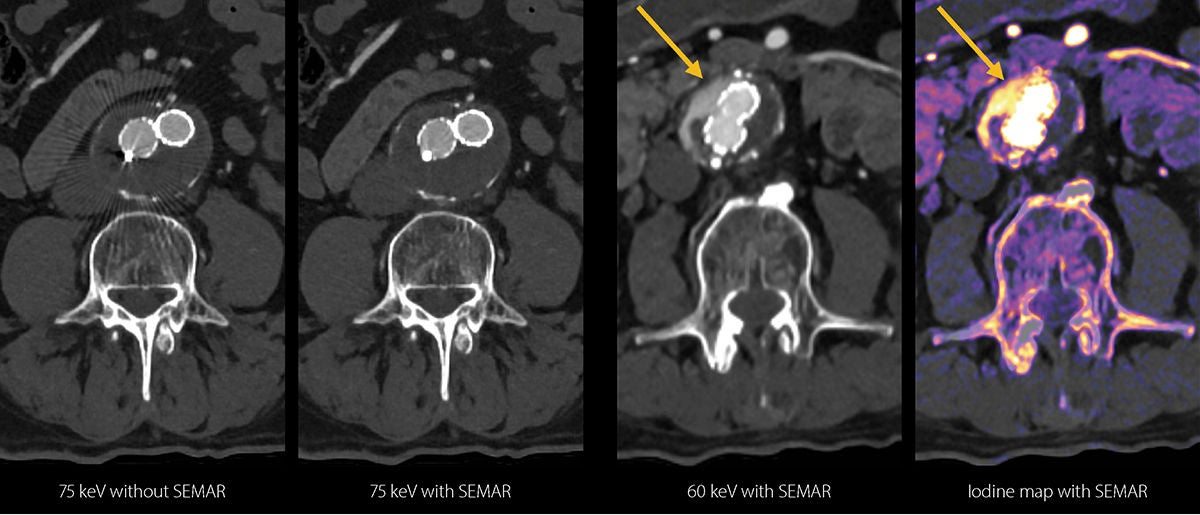 Spectral images at 75 keV without Single Energy Metal Artifact Reduction (SEMAR) demonstrate significant streak artifacts, which may preclude the assessment of endoleaks and endograft integrity. Conversely, the spectral images at 75 keV with SEMAR show excellent metal artifact reduction with improved visualization of the adjacent soft tissue. Virtual monochromatic images at 60 keV and the iodine map with SEMAR clearly demonstrate the presence of a type II endoleak (arrow). The spectral outputs combined with SEMAR improve the ability to visualize the endoleak.

“Deep Learning Spectral CTA offers state-of-the-art image quality and improves the conspicuity and characterization of arterial structures.”
“SEMAR in addition to a Spectral DLR protocol will substantially reduce metal artifacts.”
Prof. Mickaël Ohana MD, PhD
Nouvel Hôpital Civil
Strasbourg University Hospital,
Strasbourg, France.
Technology
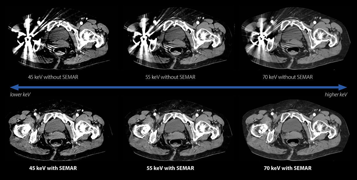
SEMAR is an innovative raw data-based reconstruction technique that can be applied to any spectral scan. Streak artifacts from metallic prostheses and endoprostheses are substantially reduced, resulting in clear visualization of adjacent anatomy.
SEMAR can be set up in the spectral scan protocol and applied automatically to all spectral reconstructions.
SEMAR can be set up in the spectral scan protocol and applied automatically to all spectral reconstructions.
Conclusion
Deep Learning Spectral CT, in combination with SEMAR, substantially reduces metal artifacts in all spectral reconstructions. This allows clinicians to fully benefit from the increased iodine conspicuity in the low keV range (35-60 keV), to identify subtle endoleaks with greater confidence. //
Social Media


