

PUREViSION Optics: The optimal balance between image quality and dose

Introduction
In CT imaging, the optimal balance between radiation dose to patients and image quality depends on many factors, including the particular clinical indication for a scan and the size of the patient. Over the years, Canon Medical has introduced a range of technologies to personalize patient dose, including automatic exposure control (AEC) systems, and later, AEC systems that could adjust to each new dose savings technology.
The evolving clinical and technological landscape has posed new challenges to optimizing the balance between image quality and dose.
For example, increasing adoption of dose-saving features, including iterative reconstruction technologies like AIDR (Adaptive Iterative Dose Reduction) 3D and FIRST (Forward projected model-based Iterative Reconstruction SoluTion), has enabled acquisition of diagnostic-quality images at very low radiation doses. In fact, iterative reconstruction has been able to generate diagnostic-quality images at doses so low that the limiting factor in going even lower became low photon counts. Maximizing the benefits of iterative reconstruction algorithms therefore required that we push this limit by developing new hardware: detector technology with the capability of converting more photons into diagnostic quality image data.
As radiation doses acceptable in clinical practice have decreased, patient populations have changed as well. Since 1975, obesity has nearly tripled worldwide, to the point where over a third of the world’s population is now overweight or obese1. Obtaining diagnostic-quality CT images on large patients poses challenges; adipose tissue preferentially absorbs low-energy X-rays, leading to imaging artifacts. A solution to optimizing imaging for patients of all sizes, by keeping doses low while maintaining diagnostic excellence, is the use of new filtration technology that optimizes the distribution of energies in the X-ray beam.
To make optimal use of new reconstruction methods and to adapt to the evolution of the global patient population, Canon Medical has developed PURE ViSION Optics, new detector and filtration technology launched on the Aquilion Prime SP, Aquilion ONE / GENESIS Edition and Aquilion Precision. The new filtration technology optimizes the X-ray beam, recalibrating the entire imaging chain. The new detector technology then takes the signal and more efficiently converts it into light. The result is an optimal balance between image quality and radiation dose. PURE ViSION Optics improves low-contrast detectability while reducing streak artifacts and noise for low-dose acquisitions. An improvement in low-contrast detectability means that physicians can more easily identify low-contrast anatomical or pathological features. A reduction in streak artifacts means better imaging of anatomical sites with high attenuation, like the shoulder, and also better imaging in the presence of metal artifacts. A reduction in noise at low doses is particularly helpful in imaging large patients and enabling the use of low-dose protocols. By optimizing the balance between image quality and dose, PURE ViSION Optics helps physicians diagnose with confidence.
With the introduction of PURE ViSION technology, we have effectively readjusted the operational range of CT to lower doses. PURE ViSION technology is only one of a variety of dose-savings features that Canon Medical has introduced, including our iterative reconstruction algorithms, AIDR 3D and FIRST; SURE Exposure automatic exposure control; and vHP (variable Helical Pitch), Canon Medical has continuously worked to provide the optimal balance between radiation dose and image quality; PURE ViSION Optics continues this tradition, enabling physicians to provide high-quality patient care.
PUREViSION Optics optimizes imaging performance
The integration of PURE ViSION Optics filtration technology with the detector’s 40% increase in light output2-4 yields measurable improvements in image quality, including:
- 12-22% improvement in low-contrast detectability for body and brain imaging at equivalent radiation dose3,4
- 13% reduction in noise at equivalent radiation dose3
- equivalent low-contrast detectability at 20-31% lower radiation dose for the body2-4
- reduced streak artifacts4
At the same radiation dose, PURE ViSION Optics yields a 12-22% improvement in low-contrast detectability3,4 for body and brain imaging2. Figure 1 illustrates PURE ViSION Optics performance on phantom images: at the same CTDI value, more of the simulated lesions on the phantom are visible on the images obtained with PURE ViSION.
At equivalent radiation dose, PURE ViSION can generate images with 13% less noise2. Figure 2 illustrates PURE ViSION’s capability to generate images with high diagnostic content and low noise. A nodule is clearly visible in the axial view on the left panel.
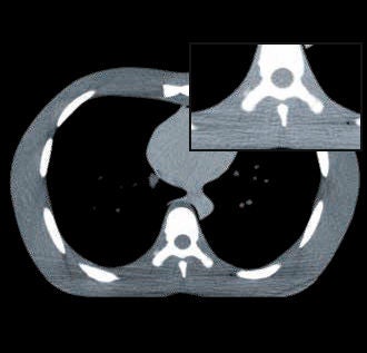
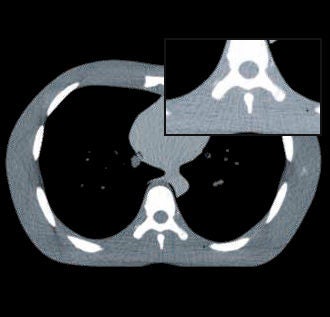
Figure 3 illustrates images of a lung phantom acquired with and without the use of PURE ViSION. Note that PURE ViSION reduces streak artifacts; the severity of streak artifacts is much less on the PURE ViSION image (right panel).
Figure 1 Regions of interest extracted from images of the Catphan® 600 phantom low-contrast module, without (left) and with (right) PUREViSION technology (Aquilion ONE on the left; Aquilion ONE / GENESIS Edition on the right). Both images were obtained at similar CTDI values (left: 58 mGy, right: 59 mGy) and reconstructed with AIDR 3D. At similar CTDI, the PUREViSION image shows improved low-contrast detectability, with more of the simulated lesions visible on the PUREViSION images.
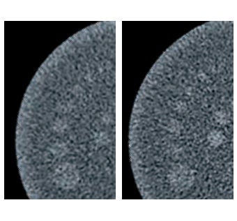
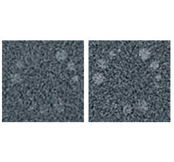
Figure 2 The introduction of AIDR 3D and FIRST iterative reconstruction has pushed imaging systems to ultra-low photon counts. This acquisition, with a nodule clearly visible in the axial view, was obtained at 2.5 CTDI, 98.9 DLP, or 1.38 mSv. PUREViSION technology reduces noise while maintaining high image quality.
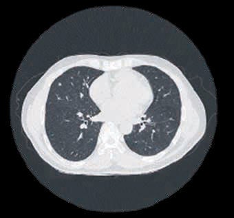
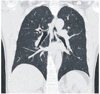
Figure 3 This image shows a lung phantom imaged with conventional (left, Aquilion ONE / ViSION Edition) and PUREViSION (right, Aquilion ONE / GENESIS Edition) technology. Streak artifact reduction is visible in the inset. These are helical scans, obtained at the following parameters: Helical, 120 kV, AEC (SD7@5 mm, FC13, AIDR 3D Standard) , 0.5 s/r , 0.5 mm × 80, HP111, 350 mmΦ, FC03, AIDR 3D OFF, CTDIvol 6.7 mGy. Display parameters: WW=300, WL=30.
PUREViSION Optics prioritizes patient safety
Canon Medical strives to provide imaging tools to generate diagnostic-quality images using the lowest achievable radiation doses. For body CT, model observer studies demonstrate that PURE ViSION yields equivalent low- contrast detectability at 20‐31% less radiation dose2. Figure 4 illustrates the dose reduction on lung images, with the image on the right obtained at approximately half the CTDI as the one on the left.
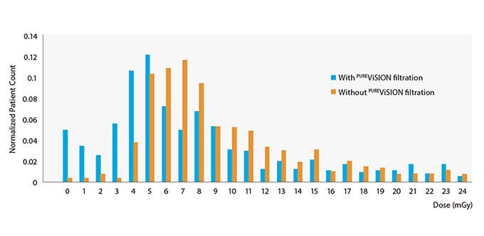
The dose reduction that PURE ViSION achieves has a demonstrable impact on patient care. Figure 5 shows the results of tracking dose on 1800 patients at Fujita Health University Hospital, the largest single medical facility in Japan. Of the patient cohort, eight hundred patients were imaged with the filtration technology that would ultimately become an element of PURE ViSION Optics. The remaining one thousand patients were imaged with conventional optics. The results show that the average dose delivered to the patients was lower with PURE ViSION filtration in place.
Figure 4 These images obtained without (left) and with (right) PUREViSION show similar image quality, at approximately half the CTDI; the left image was obtained at CTDI of 13.9 mGy; the right image at 7.2 mGy. The images were reconstructed at 5 mm slice thickness, with AIDR 3D Mild (FC 52) SD 10. Images courtesy of Fukuoka Wajiro Hospital, Japan.
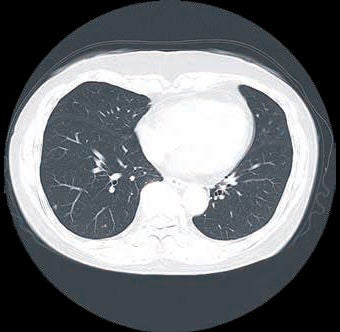
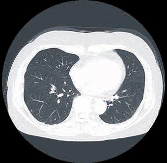
Figure 5 Frequency distribution of radiation doses delivered to 1800 patients at Fujita Health University Hospital, Japan. All patients were scanned on the Aquilion ONE / GENESIS Edition; 800 with and 1000 without the upgraded filtration technology that is now a component of our PUREViSION Optics. The new filtration technology resulted in a lower mean dose. Dose values were obtained using Vitrea™ Vitality Dose Tracking software.
PUREViSION Optics transforms routine CT imaging: A clinical case
”With PUREViSION Optics we experience a dramatic reduction in radiation dose in combination with much better image quality.”
Dr. Russell Bull, MRCP, FRCR
Consultant Radiologist, Royal Bournemouth Hospital
Bournemouth, United Kingdom
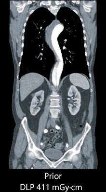
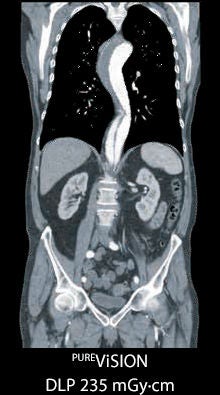
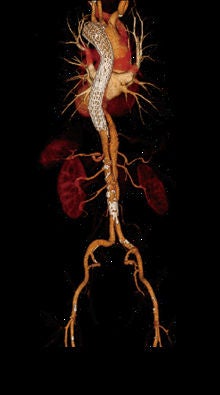
A 77-year-old man presented at the Royal Bournemouth Hospital radiology department for a follow up scan after an aorta stent placement for a type B dissection. A CTA examination of the aorta was requested to rule out complications after surgery.
The type B dissection is visible in the follow-up scan (Figure 6) two years after treatment. Contrast is visualized within the false lumen of the thoracic aorta without expansion of the lumen, indicating that the dissection is stable.
This case demonstrates the capabilities of PURE ViSION technology: The scan performed with PURE ViSION Optics technology is clearer, has fewer artifacts, and was acquired at a reduced radiation dose.
Figure 6 CTA of aorta, without (Left) and with (Right) PUREViSION Optics.
PUREViSION Optics optimizes the imaging chain
To meet the challenges of today’s clinical environment, PURE ViSION Optics optimizes each step of the imaging chain, from X-ray generation and transmission to X-ray detection.
X-ray generation: Beam shaping by PURE ViSION Optics
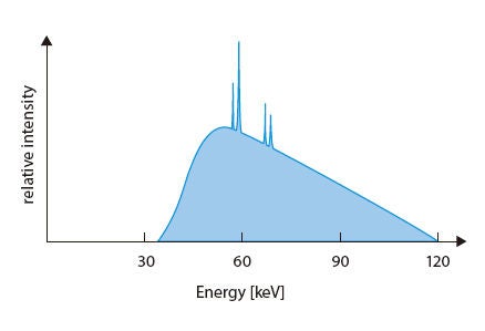
CT imaging begins with emission of radiation from the X-ray tube’s anode. The emitted X-rays are polychromatic, consisting of photons with a range of energies. Figure 7 shows a schematic of an X-ray spectrum.
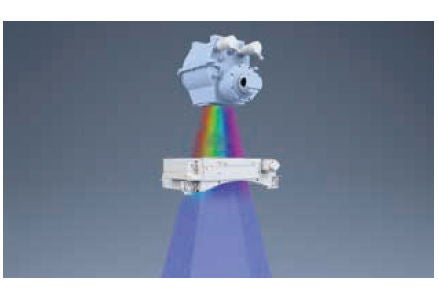
Figure 8 illustrates PURE ViSION Optics’ improved filtration, which optimizes the X-ray spectrum by removing more low-energy photons. The result is a beam with a higher effective energy, known as a “harder” beam.
Harder beams are better able to penetrate dense anatomy (such as the bony skull). Harder beams also minimize the relative radiation dose absorption of low energy, or “soft,” X-rays. Since the human body, and also iodinated contrast, preferentially absorb lower-energy, soft X-rays, these soft X-rays contribute to the radiation dose that a patient receives without contributing information to the resulting CT image. The use of a harder beam therefore enables more photons to contribute to the image. The harder beam helps optimize the balance between image quality and dose.
Because the X-ray beam becomes harder as it passes through tissue that preferentially absorbs the softer X-rays, pre-emptively removing the lower energy photons also reduces artifacts that would otherwise result from this beam hardening, potentially improving imaging of larger patients.
Although harder beams can reduce low-contrast detectability, by optimizing both filtration and detection, PURE ViSION actually enhances low-contrast detectability, as illustrated in Figure 1.
X-ray detection: PURE ViSION detector technology increases light output and decreases noise for lowdose imaging
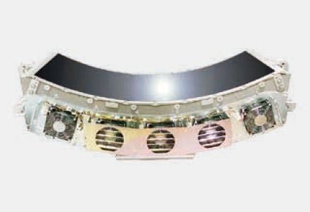
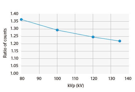
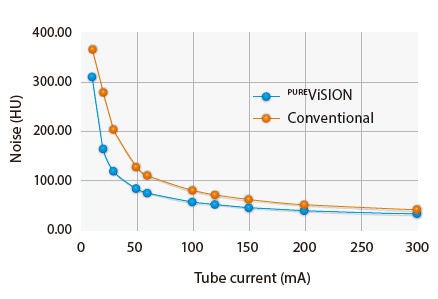
The PURE ViSION detector (Figure 9) offers 40% more light output and reduces noise. Figure 10 shows the ratio of the raw photon counts at the detector, using our upgraded filtration technology. With the upgraded filter, more photons reach the detector, which contributes to the improved light output.
Traditional detector manufacturing methods require the reassembly of detector rows into a matrix array which can result in imperfections, reducing X-ray conversion efficiency. To manufacture the PURE ViSION detector, the entire scintillator array is produced from a solid ceramic ingot. This process reduces imperfections, ensuring superior luminescent properties.
Each individual PURE ViSION detector element is delineated using precision micro-blade technology. The resulting septa (spaces between detector elements) are narrow, maximizing the surface area available for X-ray absorption. The use of microblade technology to cut the scintillator yields straight, smooth edges. The clean edges and near-microscopic septa permit more X-rays to interact with the scintillator, increasing the number of photons detected and the detection efficiency of the detectors. More scintillation means more light output and more output means more signal. In other words, rather than increasing radiation dose to increase signal, the detectors themselves avoid signal loss, capture more X-rays, and, in turn, allow for lower-noise image acquisitions.
Figure 11 demonstrates noise reduction by the PURE ViSION detector. The data are from images of a 40 cm diameter water phantom.
Canon Medical’s scientists and engineers have a history of innovation in CT detector design. Since 1999, the Aquilion CT has had the smallest detector apertures in the industry. Nearly 20 years later, Canon Medical has further improved on that record, with the introduction of the world’s first 0.25 mm aperture clinical CT detector, available in the Aquilion Precision. The PUREViSION detector is the result of many years of refining and perfecting detector design, with the goal of high quality, low-dose imaging.
Conclusions
The challenges of today’s clinical environment, which require imaging larger patients and imaging at lower doses, were the key drivers of the development of PURE ViSION Optics.
The integration of PURE ViSION Optics’ filtration technology with the detector’s 40% increase in light output3,4 yields measurable improvements in image quality, including:
- 12-22% improvement in low-contrast detectability for body and brain imaging at equivalent radiation dose3
- 13% reduction in noise at the same radiation dose3
- equivalent low-contrast detectability at 20-31% lower radiation dose2-4
- reduced streak artifacts4
PURE ViSION’s performance is apparent in clinical images, in performance testing, and most importantly, in daily use: The data in Figure 5 demonstrate a reduction in dose over a sample of 1800 patients in a hospital, showing how PURE ViSION impacted patient care and safety. PURE ViSION makes quantifiable improvements in low-contrast detectability, noise and patient dose, empowering healthcare providers to meet the challenges of today’s clinical environment and deliver the best possible patient care.
References
- World Health Organization. Obesity and overweight. World Health Organization. [Online] February 16, 2018. [Cited: September 7, 2018.] http://www.who.int/news-room/fact-sheets/detail/obesity-and-overweight.
- All results are based on measurements of detectability from a model observer study (non-prewhitening observer) on the FDA-MITA head and body phantoms. Please see “Computed Tomography Image Quality (CTIQ): Low-Contrast Detectability (LCD) Assessment When Using Dose Reduction Technology (NEMA/MITA WP 1-2017). NEMA Medical Imaging & Technology Alliance Document, July 7, 2017 for methodology.
- Aquilion Prime SP compared to Aquilion PRIME.
- Aquilion ONE / GENESIS Edition compared to Aquilion ONE.
Social Media


