

AiCE Deep Learning Reconstruction: Bringing the power of Ultra-High Resolution CT to routine imaging

Introduction
The state-of-the-art in the field of Artificial Intelligence (AI) is a subfield of machine learning known as deep learning. Deep learning takes advantage of multilayered artificial neural networks to produce results that have taken the AI world by storm—outperforming even humans at tasks such as object recognition. In fact, deep learning has opened the door to a host of applications that are solving complex problems in our daily lives, from the navigation systems in our cars, to near-instant language translation applications on our phones, to automatic photo recognition and labelling on social media and much more. Unlike conventional algorithms that are constrained by pre-programmed rules for performing a complex task, deep learning occurs when a neural network learns from its own intensive training process and develops its own logic structure. Canon Medical is proud to introduce the AiCE (Advanced Intelligent Clear-IQ Engine), Deep Learning Reconstruction (DLR) algorithm for CT (Computed Tomography), featuring a deep learning neural network that can differentiate and remove noise from signal, creating extraordinary high quality images.
The goal of image reconstruction in CT is to facilitate the diagnosis of the patient by converting the raw projection data into an image of the highest possible quality. Prior to DLR, a reconstruction algorithm that produced exceptional low contrast detectability, preserved spatial resolution, and markedly reduced noise and artifact—without requiring an increase in radiation dose or hindering workflow—was a tool that remained elusive. Even the best Model-Based Iterative Reconstruction (MBIR) algorithms can suffer from poor noise texture at low dose levels and reconstruction times that are workflow-prohibitive in a busy clinical environment. Canon Medical’s introduction of the Ultra-High Resolution Aquilion Precision, capable of 150 μm × 150 μm × 200 μm resolution, further motivated the need for development of next generation reconstruction technology—a fast, low noise algorithm able to preserve an extraordinary level of detail. The result is AiCE, a fully-integrated DLR that not only preserves the Precision’s extraordinary spatial resolution but also simultaneously improves noise and low contrast characteristics. With AiCE dose neutrality is achieved between Precision’s Ultra-High Resolution scan modes vs conventional resolution scanning reconstructed with traditional hybrid iterative reconstruction. Together, AiCE and Precision bring the power of deep learning and Ultra-High Resolution scanning at standard radiation doses to everyday use in the clinic, putting the future of CT in your hands.
Aquilion Precision and AiCE DLR
The Aquilion Precision was designed to visualize anatomy and pathology in routine imaging with double the level of detail of conventional resolution systems. The first major step in achieving such a fine degree of detail with the Aquilion Precision was the invention of a 0.25 mm × 160 detector1. The Precision detector is crafted with proprietary cutting techniques that generate discrete, optically-isolated detector elements that allow for ultrathin septa which permit a substantial increase in light-sensitive area on each element with minimal crosstalk. This advancement, coupled with innovations in scintillator efficiency, detector circuitry and other DAS components, has led to the most dose efficient detector in Canon Medical history. The second key step in creating the Ultra-High Resolution Precision was a new tube system, featuring reduced focal spot sizes, as small 0.4 mm × 0.5 mm and rotating at 10,000 rpm to efficiently dissipate heat. And, now, the third and final critical step toward routine Ultra-High Resolution CT is AiCE DLR, a fast reconstruction algorithm including both raw data and image domain components to reduce artifact and improve the signal-to-noise ratio. The AiCE DLR features a highly-trained, multilayer neural network to reduce the magnitude of noise in high resolution images while preserving Precision’s detail. The combination of Precision with AiCE DLR allows for Ultra-High Resolution scanning at standard clinical CT doses for the first time.
Workflow Efficiency: Achieving the speed you need in a busy clinical environment
During engineering development, the AiCE DLR algorithm is taught to produce high signal-to-noise ratio (SNR) images through an intense training process. AiCE learns to differentiate signal from noise by training on select, high quality patient data sets acquired with high tube current and reconstructed with all the benefits of state-of-the-art MBIR—including sophisticated system and noise models as well as a large number of iterations not possible clinically. Because this time-consuming training process is completed before leaving the factory, the fully-trained AiCE DLR is able to work quickly in the clinic, reconstructing an Ultra-High Resolution 1024 × 1024 abdominal case acquired with 0.25 mm slices quickly. This rapid reconstruction allows the clinician to take advantage of the benefits of deep learning, which by design incorporates the all sophisticated modelling utilized in MBIR, in a time-efficient manner: working at over five times the speed of MBIR reconstruction, AiCE DLR is making the benefits of low noise, Ultra-High Resolution an everyday reality.
Like Canon Medical’s iterative reconstruction algorithms, AiCE is also fully integrated into the SURE Exposure mA modulation system. The system automatically adjusts each individual patient’s mA profile based on the associated benefits and dose reduction abilities of AiCE reconstruction. AiCE DLR is available in three straightforward settings, Mild, Standard, and Strong, making the application of AiCE simple and easy-to-use.
Low Contrast Detectability: Dose neutral ultra-high spatial resolution
AiCE DLR is able to dramatically decrease the magnitude of noise in an image, improving low contrast detectability. When used with the Aquilion Precision’s Ultra-High Resolution mode, which acquires data with a 0.25 mm nominal slice width and reconstructs to 1024 × 1024 matrix, AiCE DLR can achieve the same low contrast detectability as conventional resolution images reconstructed with AIDR (Adaptive Iterative Dose Reduction) 3D. This combination of technologies opens up the advantages of Ultra-High Resolution imaging to the clinician without concern of low contrast detectability loss or increased radiation dose to the patient.
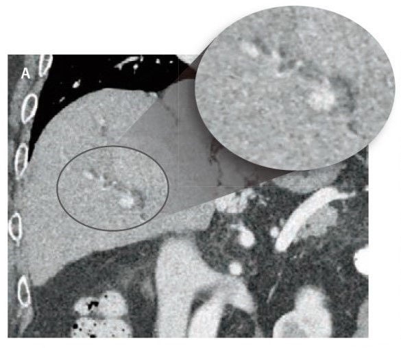
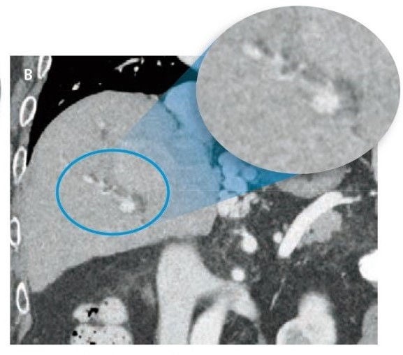
Notice the visibility of the lesions on the Ultra-High Resolution image reconstruction AiCE at 12.4 mGy compared to the normal resolution image reconstructed with AIDR 3D at 11.8 mGy in Figure 1. Figure 2 compares a normal resolution AIDR 3D phantom image with an Ultra- High Resolution AiCE phantom image at the same dose.
In addition, AiCE DLR reduces the magnitude of visually unappealing noise textures found at low dose with iterative reconstruction algorithms, resulting in an appearance more similar to low noise filtered backprojection as show in Figure 3.
Figure 1 Improved liver lesion visibility on UHR CT images with similar radiation dose. AIDR 3D at 11.8mGy (A) compared to AiCE at 12.4 mGy (B).
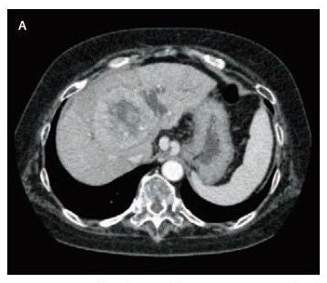
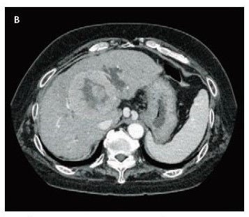
Figure 2 Catphan® LCD module acquired with normal resolution mode, reconstructed with AIDR3D (A) and acquired in Ultra-High Resolution mode, reconstructed with AiCE (B).
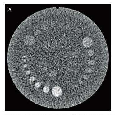
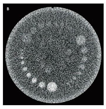
Figure 3 Improved noise properties on Ultra-High Resolution abdominal images with AiCE. A: MBIR, B: AiCE
Spatial Resolution
Because AiCE is trained on images of the highest quality using MBIR reconstruction, AiCE DLR learns to preserve edge and maintain image detail, which is particularly important for Ultra-High Resolution scanning. The Aquilion Precision has a 0.25 mm × 160 detector and is paired with a new X-ray tube design, featuring reduced focal spot sizes, as small 0.4 mm × 0.5 mm, resulting in twice the spatial resolution of conventional systems.
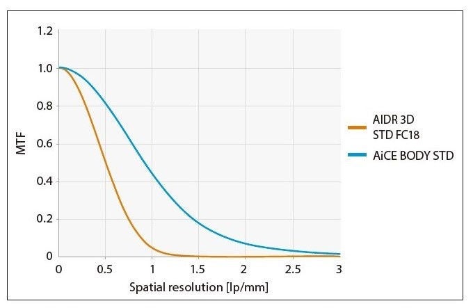
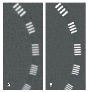
Because it incorporates the spatial resolution benefits of MBIR, AiCE improves high contrast resolution compared to hybrid iterative reconstruction techniques such as AIDR 3D. This spatial resolution improvement is demonstrated in the Modulation Transfer Functions (MTF) in Figure 4. This spatial resolution improvement facilitates the visualization of fine detail, such as vasculature and fine pathology illustrated in Figure 5 using the line pair phantom.
Looking to the future with the Precision CT and AiCE DLR
Ultra-High Resolution scanning combined with DLR will continue to expand the role of CT in the diagnosis of a wide assortment of clinical presentations. Clinical applications such as lung, temporal bone, vasculature, stent structure, artifact reduction, as well as visualization of small tumors and structures all have potential to benefit from Precision with AiCE DLR. In addition, the burgeoning discipline of radionomics, a field of medical study that aims to extract large amount of quantitative features, such as shape, size, and texture, from medical images using data-characterization algorithms, is expected to benefit greatly from Precision with DLR—potentially leading to advances in decision support, precision medicine, population health, prognosis assessment and predicted treatment response.
AiCE DLR: How Does it All Work?
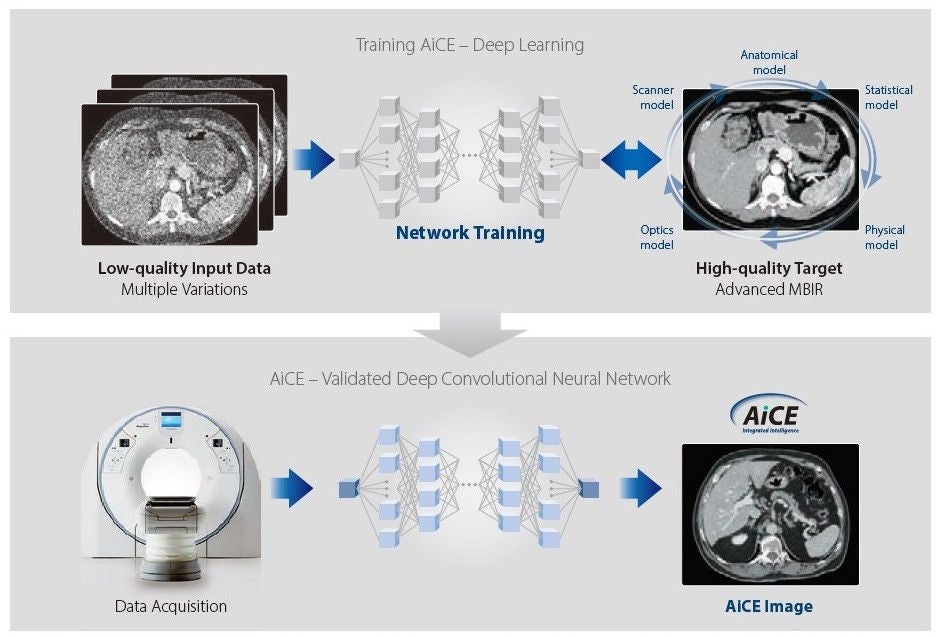
Figure 6 Overview of AiCE Deep Learning Reconstruction: The AiCE DLR is Trained with high quality, advanced MBIR Target Images and learns to turn low quality input data into low noise images that are sharp and clear. In the clinic, AiCE DLR operates in the raw and image domain to efficiently reconstruct images.
Deep Learning Overview
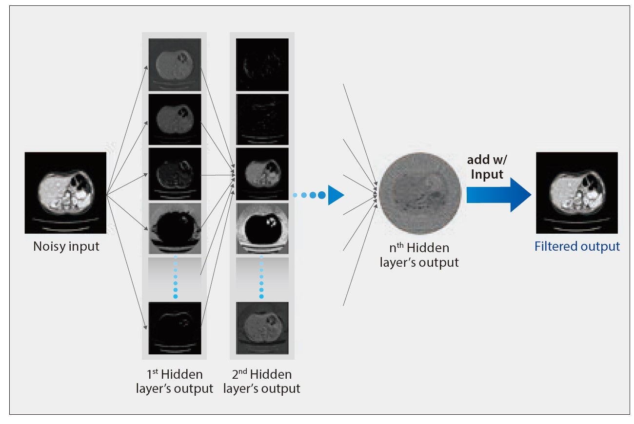
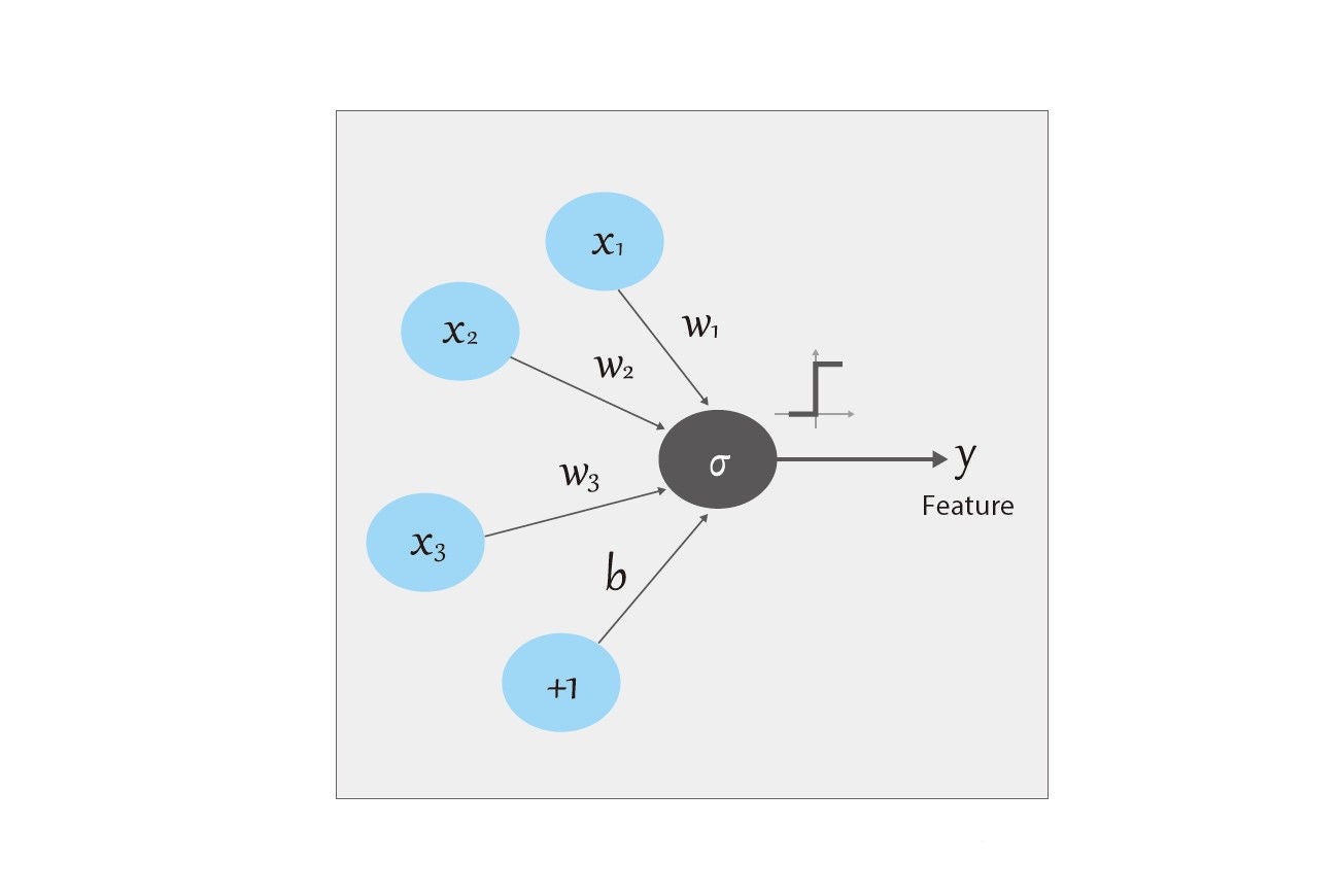
Deep learning, the latest in AI, has been successfully applied to such tasks as image recognition, segmentation and classification. With deep learning, a Deep Convolutional Neural Network (DCNN) comprised of layers of neurons is trained in the performance of a complex task. A neuron, illustrated in Figure 8, is a node where a mathematical operation takes place, the output of which is connected with other neurons, forming a network. The neural network derives its name from the neuron-synapse paradigm found in biology and mimics how humans draw conclusions, based on learning from examples. This ability to learn via a deep neural network gives the deep learning algorithm the freedom to find the optimum way to perform the desired task. DCNNs have shown extraordinary performance at image classification tasks, bursting onto the artificial intelligence scene in 2012 by winning the ImageNet challenge—skillfully classifying one thousand object types from over one million images. A basic DCNN structure is illustrated in Figure 7.
AiCE DLR: Training
The key to a successful DCNN lies in its training, the process by which the DLR learns how to successfully perform its function. The network must compare its output image to a gold standard reference image in order to gauge its performance and learn, i.e. adjust the weights of its neurons. In order to do this the DCNN uses a mathematical loss function to determine the amount of error between its output and the reference datasets. In the case of AiCE, the gold standard clinical reference images are acquired with high tube current and reconstructed with true MBIR reconstruction, which takes into account modelling of the system optics, system physics, scanner statistical properties and human anatomy, and uses a greater number of iterations than could be otherwise used in a clinical setting due to time constraints. Using the estimation of error between the output of the DCNN and the gold standard, the DCNN the error estimate through the network and adjustments are made to the neuron’s weights in order to reduce the discrepancy. This input-forward, error-backpropagation process is iteratively repeated until the network is optimized. In order to ensure optimal results, millions of image pairs were used in the training of AiCE DLR. This complex training process is completed in development with no off-site unsupervised training, which could alter algorithm performance, taking place.
In order to make sure AiCE DLR is robust in low dose situations AiCE’s training included low quality data sets, used to teach AiCE how generate high quality images from low quality images while preserving signal and spatial resolution across the clinical spectrum. The algorithm was tested with independent validation datasets, to ensure wide applicability of the algorithm and avoid a phenomenon in machine learning known as overfitting, which occurs when an algorithm is too finely tuned to the training data to be robust against new inputs. Thousands of phantom and patient images were examined by medical physicists and radiologists in the development of the AiCE DLR reconstruction algorithm.
Inside AiCE DLR
The AiCE DLR applies the extraordinary classification abilities of a DCNN to the task of differentiating noise and signal in CT images and enhancing signal while suppressing noise to generate a high quality image for clinician interpretation. An overview of the AiCE DLR process is given in Figure 6. The AiCE reconstruction process begins in the raw data domain where AiCE analyzes the raw data and, armed with detailed scanner model information, makes modifications. These modifications in the projection domain improve output SNR and reduce artifacts, such as streaks. This raw data is then initially reconstructed to form a seed image, known as the “input layer”, to the DCNN.
Once the input image is fed into the DCNN, it is analyzed by several network layers referred to as “hidden layers.” The hidden layers of a DCNN contain convolutional layers, in which the component neurons act as feature selectors on small patches of data. In a traditional heuristic algorithm explicit image features, such as a curved edge, would be pre-selected by the programmer and “convolved,” i.e., filtered, with the image data. During the deep learning process, each neuron in a convolutional layer learns what features to look for based on the training data. AiCE’s DCNN has thousands of neurons, thoroughly sampling feature space. The network “learns” image features and their level of importance by adjusting the parameters, known as weight and bias, utilized by each neuron in the convolutional layer.
The output of the convolutional layer is the fed into an “activation layer.” In biology, a neuron only fires when the input to it surpasses a threshold. Similarly, the activation layer in a DCNN serves an analogous purpose in that, based on the strength of a neuronal response to the input data, the activation layer determines which neuron responses will pass to the next layer in the DCNN. After passing through all the hidden layers of the AICE neural network, the signal and noise are separated and a signal image, known as the output layer, is generated for the user.
One key to a successful DCNN lies in its network structure design, which impacts both image quality and reconstruction speed. To achieve the best computational efficiency and improve output image quality, network structure factors such as number of network layers, number of neurons in each layer, convolution kernel sizes, etc, were fully optimized in the AiCE algorithm. Elegant acceleration strategies and memory management technologies were carefully designed and integrated in the system to fully utilize hardware capabilities and maximize reconstruction speed.
Putting the future in your hands
Integrated, effective, and easy-to-use, AiCE DLR brings the power of deep learning to the world of Ultra-High Resolution CT. The raw data domain aspects of AiCE DLR combined with the ability of the deep convolutional neural network to differentiate signal and noise leads to a wealth of advantages compared to other forms of reconstruction, including improvements in noise, low contrast detectability, and spatial resolution preservation—making Ultra-High Resolution CT at standard doses available to the clinician. The combination of the Aquilion Precision and AiCE DLR provides new opportunities for advancement in patient diagnosis, clinical applications, as well as radionomics and represents the future of computed tomography.
References:
1. Boedeker K., Aquilion Precision Ultra-High Resolution CT:
Quantifying diagnostic image quality, Canon Medical Systems Corporation, 2017
Social Media


