

New & Emerging Possibilities Using Angio 4D CT in IR
– The University of Chicago Medicine
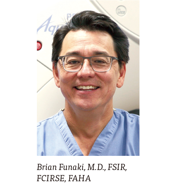
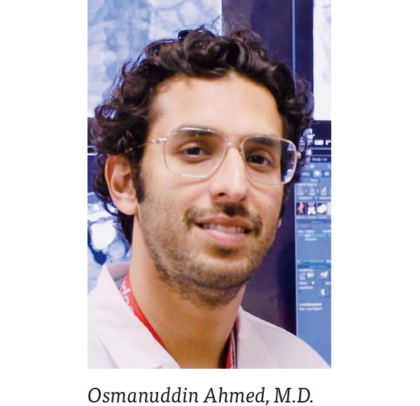
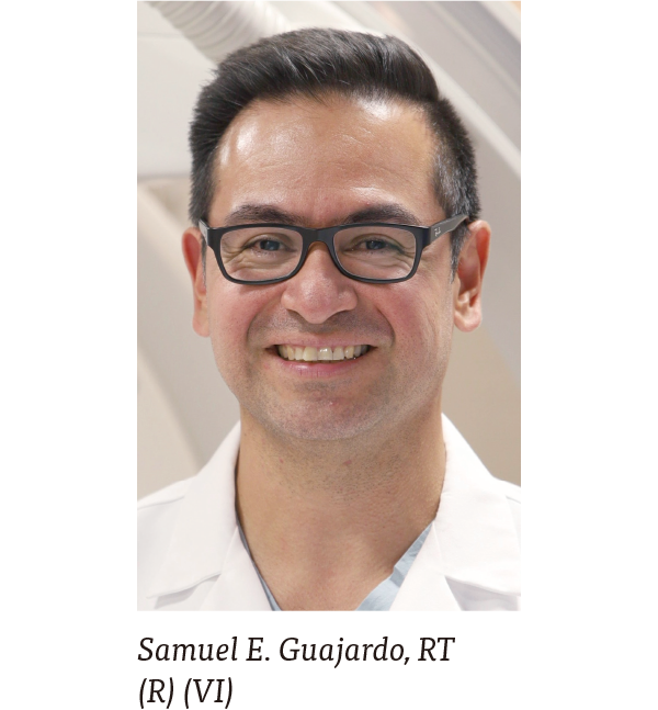
Brian Funaki, M.D., FSIR, FCIRSE, FAHA, Osmanuddin Ahmed, M.D., Rakesh Navuluri, M.D., Steven M. Zangan, M.D., Samuel E. Guajardo, RT (R) (VI)
Department of Radiology, Section of Vascular and Interventional Radiology University of Chicago Medicine, Chicago, IL, USA
Betty Ashdown A.R.R.T. (R) (CV) (CT) RCIS, FAVIR, FSICP, Canon Medical Systems USA, Inc. Strategic Development Manager
Yiemeng Hoi, Ph.D., Canon Medical Systems USA, Inc., Medical Affairs Manager
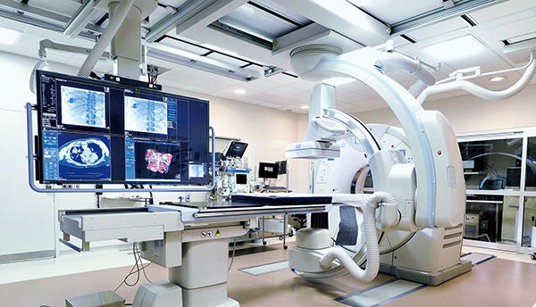
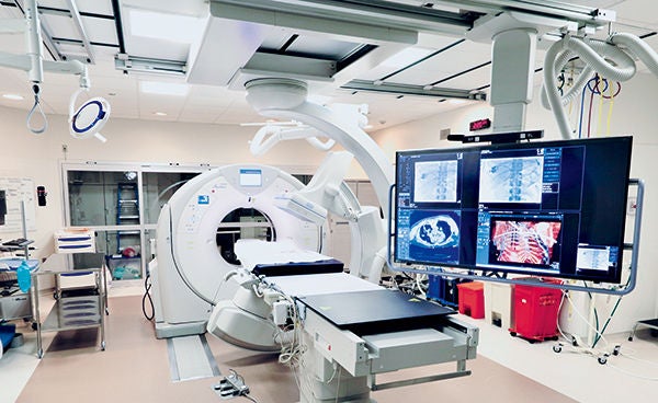 University of Chicago 4D CT PRIME system (Photo Credit: Mark Hohn)
Better patient care in one Interventional Lab/CT solution with Canon Medical’s 4D CT
Canon Medical Systems’ 4D CT is a powerful hybrid imaging system which has a fully operational ceiling mounted angiography system combined with an advanced dynamic volume CT scanner. This combination in one suite delivers an exceptional solution for more complex image-guided interventional procedures. The 4D CT has a specially designed extendable table top that easily slides into position during imaging for either the angiography C-arm or the CT scanner. The uniqueness of the 4D CT is the ability of clinicians to prioritize the patient experience and streamline their workflow during interventional procedures in a single clinical setting. The combination of both systems within one integrated imaging suite enables physicians to eliminate patient transfer during intricate procedures and confirm the effectiveness of the procedure.
The Canon Medical 4D CT ceiling mounted C-arm has a high resolution flat panel detector with compact housing to enable unprecedented flexibility of positioning the C-arm 270 degrees around the patient. The CT system offers low contrast detectability and high contrast resolution for either volume or helical examinations to create thin slices with high precision 3D and MPR images.
The history of the University of Chicago Medicine dates back to 1927 and is a not-for-profit academic medical health system located on the campus of the University of Chicago in Hyde Park. The Department of Radiology, Vascular and Interventional Radiology team installed Canon Medical’s 4D CT in April 2018. The physicians have learned the capabilities of the system and the various types of procedures performed continue to grow.
The procedures in this article are due to the efforts of the physicians and staff at the University of Chicago and is intended to highlight countless categories of procedures that are being performed.
Renal Aneurysm Embolization
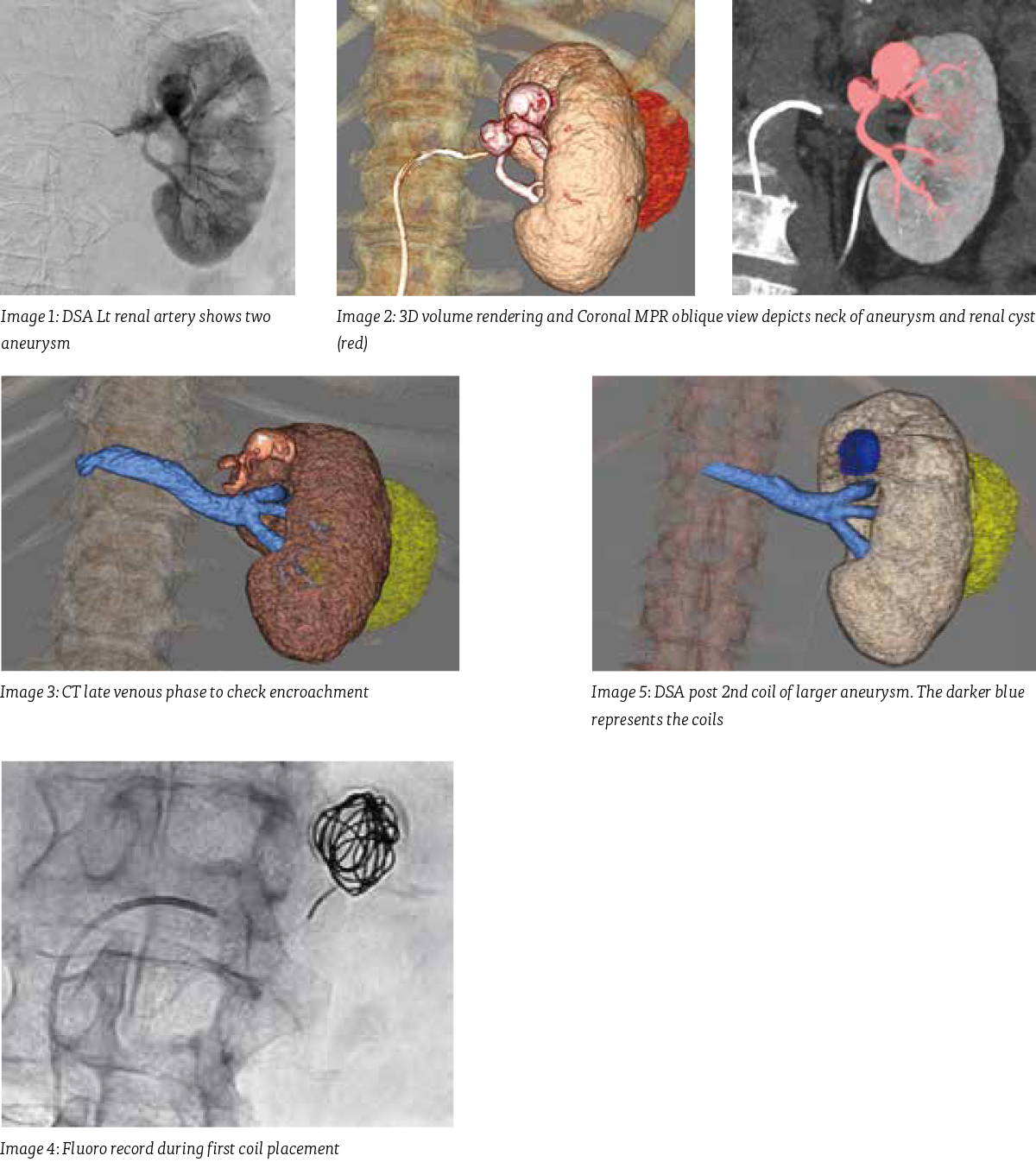
71 year old man with a past history of esophageal cancer had two renal aneurysms discovered incidentally on abdominal CT performed to evaluate tumor response. The smaller one was 1.8 cm and the larger one 3.6 cm in diameter. Initial DSA (digital subtraction angiography) series performed at 3 frames per second (fps) showed two aneurysms originating off of the Lt renal artery as well as a large cyst. The neck of the aneurysms were not delineated by DSA alone so a helical CT scan was performed with an intra-arterial injection of the Lt renal artery. The multiplanar views obtained from the CT aided in localizing the origin of the neck of both aneurysms. An additional CT scan was then performed using delayed scanning of 6 seconds to capture the left renal vein to confirm the aneurysms did not encroach on the vein. The larger one was treated using 2 long detachable packing coils to completely fill the aneurysm with preservation of all vessels. Post coiling both a DSA and a CT scan were done to check the effectiveness of the coils. The smaller aneurysm was not treated during this procedure.
Yttrium-90 Mapping
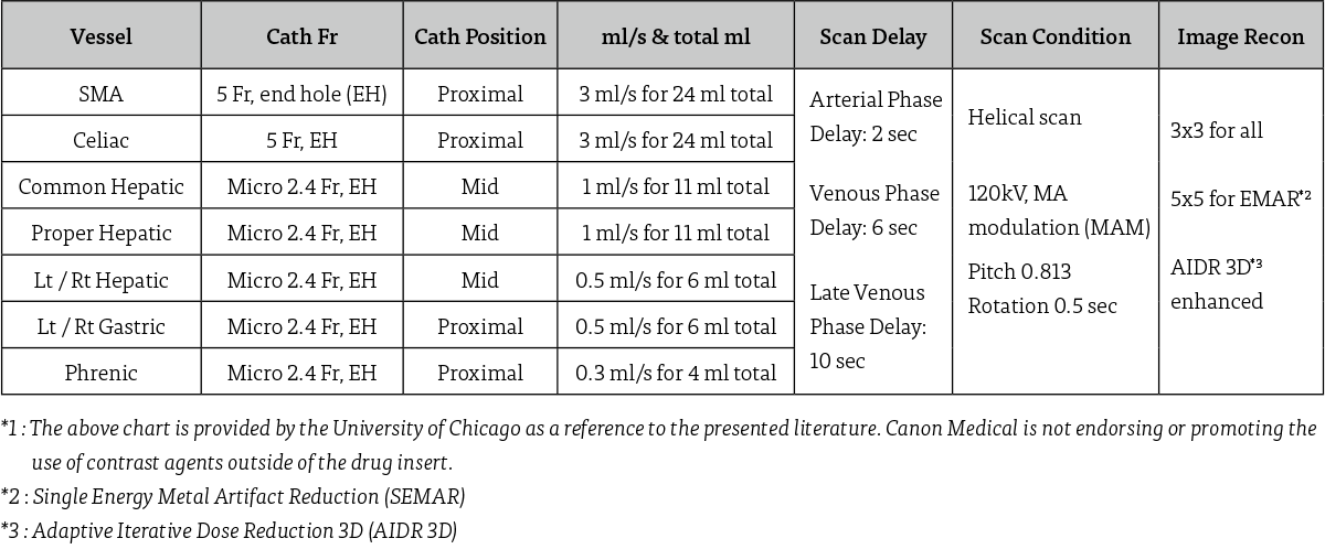
A 64 year old with a history of metastatic colon cancer in the liver, was brought to the 4D CT room for Y-90 mapping. A 5 French catheter was inserted into the patient’s right femoral artery and advanced to the celiac artery. An intraarterial injection DSA did not highlight the common hepatic artery. The common hepatic artery was found as a separate branch off of the aorta. A DSA through a 5 French catheter showed the vessels which were supplying the tumor in the late venous phase. A 2.4 French microcatheter was then advanced to the common hepatic artery and a DSA was done with an injection of 1 ml per second for a total of 11 ml.
To highlight the tumor, a tri-phasic CT scan was done with an intra-arterial injection through the 2.4 French microcatheter to identify a significant lesion in the upper right lobe of the liver. Contrast injection of 0.5 ml per second for a total of 6 ml was used with a 2 second scan delay for the arterial phase, a 6 second scan delay for the early venous phase, and then a 10 second scan delay for the late venous phase.
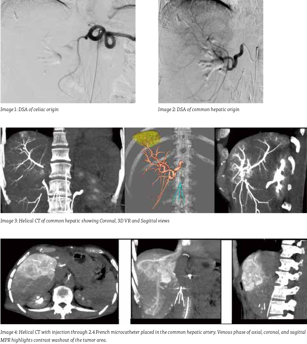
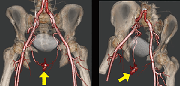 Image 1: CT of Aortic bifurcation. 3D Volume rendered AP and Oblique views
Arterial Bleed Embolization
A 23 year old man that came into the emergency room after a straddle injury and high flow priapism which developed post injury. Patient was brought to the 4D CT room and CT scan identified an arterial cavernous fistula from the right internal Pudendal artery. The communication was embolized using two microcoils and his condition resolved over the subsequent week.
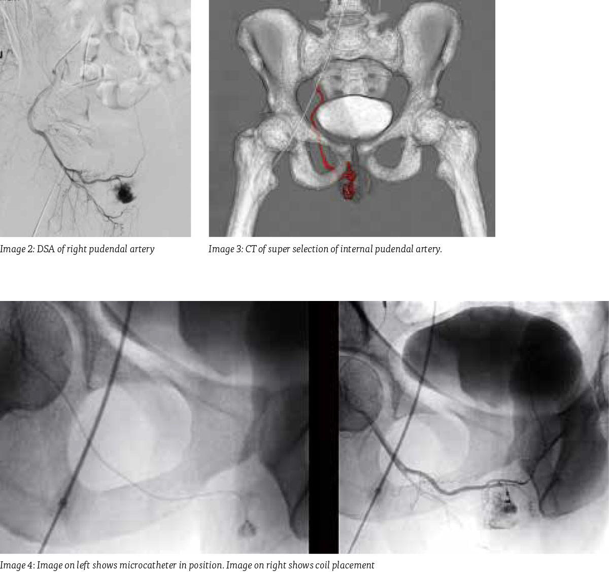
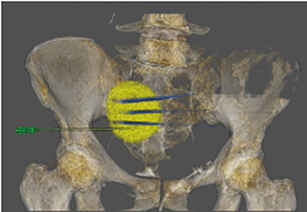 Image 1: Depicting CT-guided positioning of the cryoprobes with ice ball development.
Pelvic Mass Cryoablation With Biopsy
A 55 year old brought into the 4D CT room for possible cryoablation in the pelvis. The helical CT scan of the pelvis was done and a large mass was seen. A biopsy was necessary to specify what category of pathology. Once the biopsy needle was advanced to the mass, a CT scan was done with an intra venous injection to ensure that the needle had missed the arteries in the pelvis. A cryoablation of the mass was then performed.
Three cryoprobes were inserted into the area mass. CT provided the necessary information during this procedure without the need for fluoroscopy.The patient was able to have both procedures in the same room, on the same day without being transferred from one room to another.
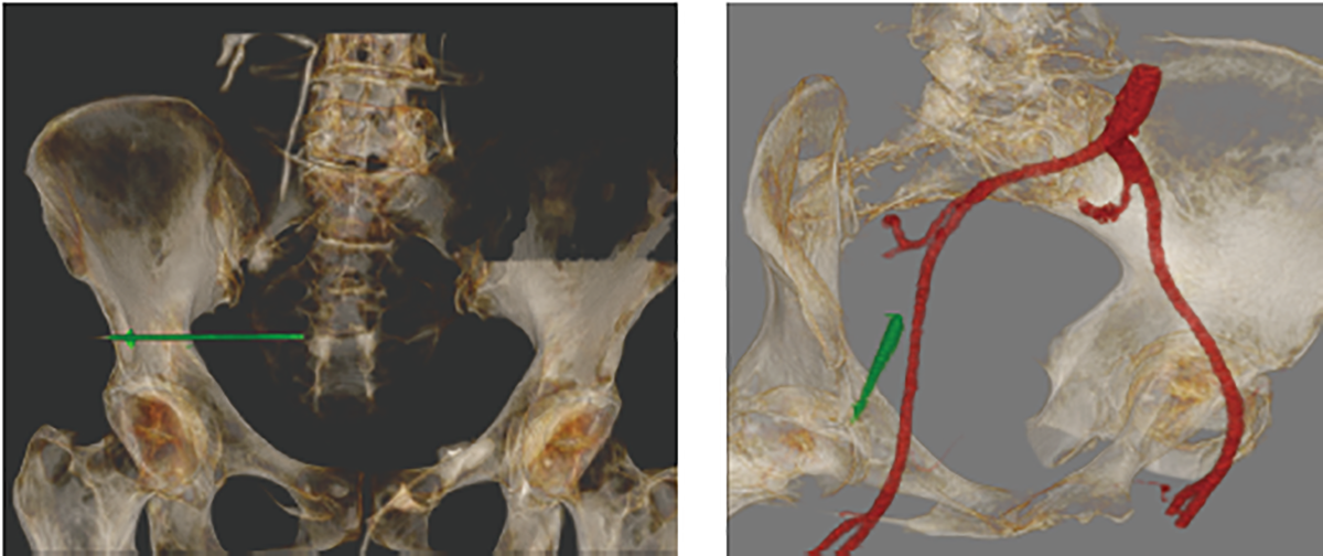 Image 2: Image on left shows the biopsy needle insertion. Image on right is the CT with IV contrast injection to check the needle pathway
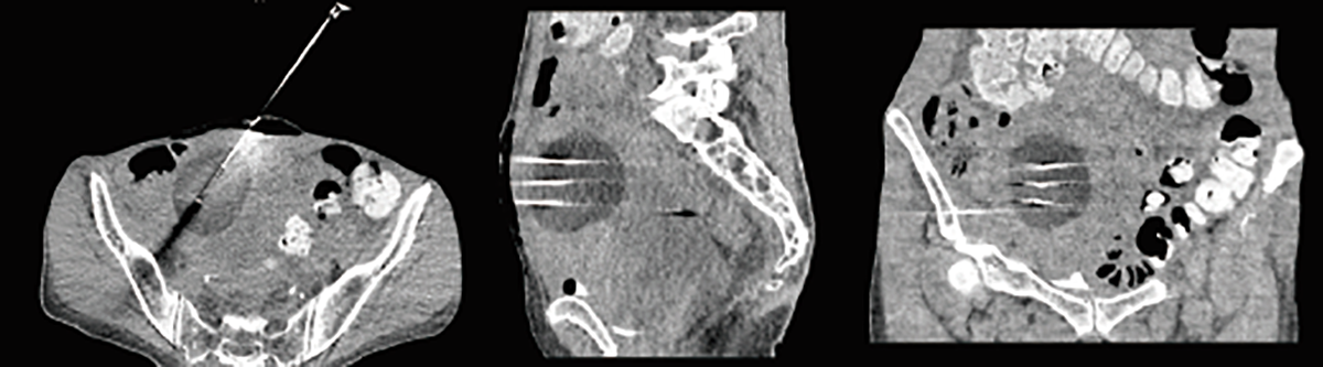 Image 3: CT scan shows axial, sagittal, and coronal MPR views of the probes for cryoablation
Type 2 Endoleak Repair
A 72 year old who had undergone an endovascular aortic aneurysm repair (EVAR). Physician suspected an endoleak outside lumen of the patient’s endoluminal stent graft. The procedure was done in the 4D CT room in anticipation of the need to perform CT scanning for localization of the area of the leak. A 5 French catheter was inserted into the right femoral artery and a DSA of the aorta was obtained.
A helical CT scan was then performed and 3D volume rendered image was used along with the MPR views to try to visualize the endoleak. The patient was found to have a Type 2 endoleak demonstrated by a dynamic CTA. The sac was accessed in retrograde fashion from the SMA into ascending left colic and then the inferior mesenteric artery. The sac and IMA ostium was embolized using microcoils.
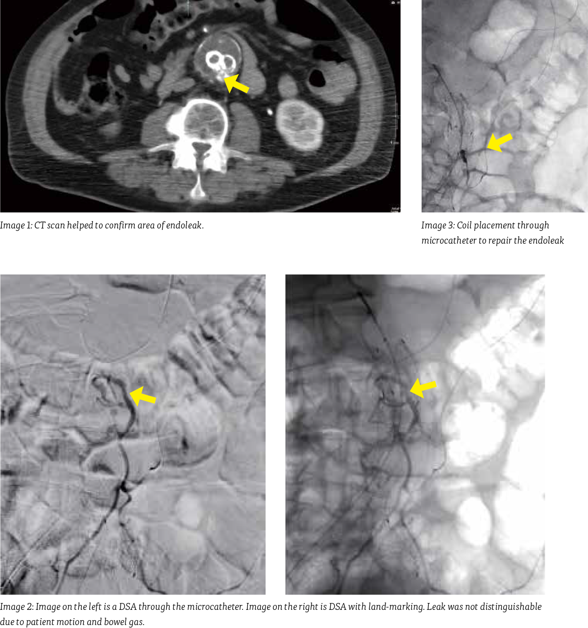
Procedural Parameters*1
Due to the amount of procedures the University of Chicago has performed and the number of staff that need to be trained, Sam Guajardo has collected information for intra-arterial injections during CT scanning that are used consistently. The following parameters are used for intra-arterial injections acquired with CT imaging
 University of Chicago 4D CT PRIME system (Photo Credit: Mark Hohn)
Conclusion
The Canon Medical 4D CT efficiently integrates CT and IR imaging into one seamless solution. The use of real CT imaging available on demand helps reduce motion and breathing artifacts due to short acquisition times and provides enhanced soft tissue visualization. The 4D CT seamlessly integrates our flexible interventional system with the advanced Aquilion CT imaging suite into one versatile solution. With the ability to see, diagnose, plan, treat, and verify in the same room, the 4D CT helps you prioritize safety, speed, and efficiently during complex interventions.Physicians are envisioning more and more ways to use the 4D CT system, while being able to change the treatment of the patients and provide a more comfortable environment during interventional procedures. //
Social Media


