

Expanding the potential of IR with Canon Medical’s Alphenix 4D CT
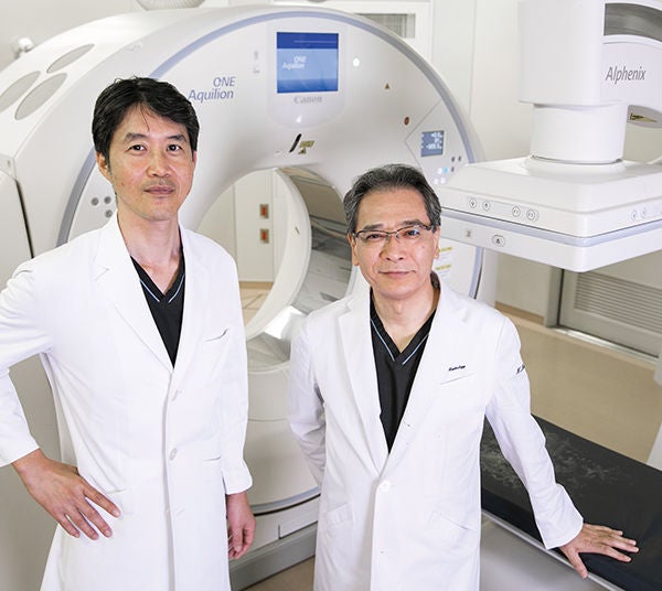 Toshihiro Tanaka, M.D., Ph.D. Associate Professor, Dept. of Radiology, IVR Center Nara Medical University / Kimihiko Kichikawa, M.D. Vice-President of Nara Medical University Hospital Professor & Chairman of Dept. of Radiology Director of Interventional Radiology Center, Nara Medical University President of Japanese Society of Interventional Radiology (JSIR)
The Department of Radiology at Nara Medical University has always been on the cutting-edge of interventional radiology (IR). As it stands, they perform more than 1,500 IR procedures annually, covering virtually all areas except for the cardiac region. Three angiography systems are currently in operation in the Department of Radiology, but only one of these systems incorporates a CT scanner: the Alphenix 4D CT system produced by Canon Medical Systems Corporation, Japan.
This system was installed in March 2019 as a replacement for their Infinix-i 4D CT system (incorporating a 16-row CT scanner) which had been in use for 16 years. To meet the needs of the rapidly expanding field of IR, they wanted a new system that could provide wider single-scan coverage, improved image quality, and reduced dose.
This system was installed in March 2019 as a replacement for their Infinix-i 4D CT system (incorporating a 16-row CT scanner) which had been in use for 16 years. To meet the needs of the rapidly expanding field of IR, they wanted a new system that could provide wider single-scan coverage, improved image quality, and reduced dose.
We recently sat down with Dr. Kimihiko Kichikawa (a Professor in the Department of Radiology) and Dr. Toshihiro Tanaka (an Associate Professor in the same department) to find out more about their experience with the Alphenix 4D CT system and how they expect the relationship between hybrid angiography-CT and IR to evolve.
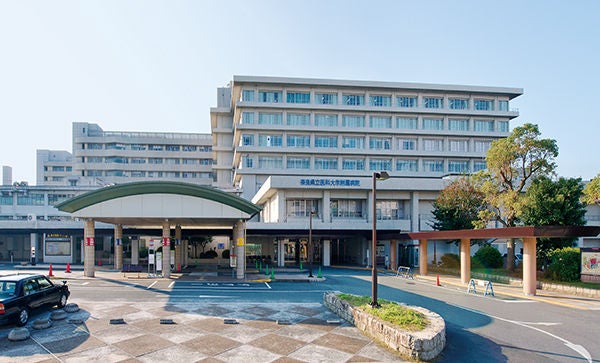 Nara Medical University
Q1: Why did you choose the Alphenix 4D CT system?
Dr. Kichikawa: We chose the Alphenix 4D CT for a couple of reasons. Firstly, it is an extremely dynamic, high-performance system that features the largest area detector available. The importance of this cannot be underestimated as it enables us to acquire volumetric data over a range of 16 cm. Secondly, 4D CT data is changing everything in the IR space. Not only does it help us decide whether or not IR is a reasonable therapeutic option, it also helps us create effective treatment plans that incorporate the right therapeutic strategy. In addition, Dynamic Volume CT helps us to obtain a clearer understanding of blood flow dynamics, with a particular focus on the feeding vessels.
Dr. Kichikawa: We chose the Alphenix 4D CT for a couple of reasons. Firstly, it is an extremely dynamic, high-performance system that features the largest area detector available. The importance of this cannot be underestimated as it enables us to acquire volumetric data over a range of 16 cm. Secondly, 4D CT data is changing everything in the IR space. Not only does it help us decide whether or not IR is a reasonable therapeutic option, it also helps us create effective treatment plans that incorporate the right therapeutic strategy. In addition, Dynamic Volume CT helps us to obtain a clearer understanding of blood flow dynamics, with a particular focus on the feeding vessels.
Dr. Tanaka: With the Alphenix 4D CT system, conventional 3D imaging is far superior with higher scanning speeds and a quicker display of reconstructed images. Different types of reconstructed images are available, and the system allows us to select the most suitable reconstruction for a range of cases, including those that require immediate confirmation or a more detailed perspective. The 4D CT is especially effective in vascular IR for patients with fast blood flow in whom we need to obtain the required information without delay. It has also proven to be a significant advance in non-vascular IR because it allows us to introduce the needle at a craniocaudal angle during puncture procedures. This means that we can now match a specific reconstruction to the clinical needs of each patient. For example, when we require highly detailed images, we can use Dynamic Volume CT, and when we simply need to scan quickly in order to view images while performing a procedure, we can use the low-dose cone-beam CT.
Q2: What changes have you seen since installing the Alphenix 4D CT system?
Dr. Kichikawa: The hybrid angiography-CT solution is essential for identifying tumor vessels and determining the extent of embolization in transcatheter arterial embolization procedures. In particular, we have found the Alphenix 4D CT system to be indispensable when performing extremely fine and selective arterial embolization, a method that was first introduced and is currently being refined by Dr. Tanaka.
Dr. Tanaka: In patients with complicated blood flow dynamics, such as those with an arteriovenous malformation or arterial dissection, we first obtain 4D CT data and carefully evaluate the reconstructed images. Next, we decide on the optimal therapeutic strategy before commencing the procedure. When we use a needle to drain an abscess, Dynamic Volume CT is an essential tool for obtaining clear images that are free of artifacts, allowing us to clearly visualize the position of the needle relative to surrounding structures. In my experience, there are serious limitations when such procedures are performed using only cone-beam CT. Alphenix 4D CT has made it possible to formulate precise therapeutic plans and to ensure the accurate implementation of IR.
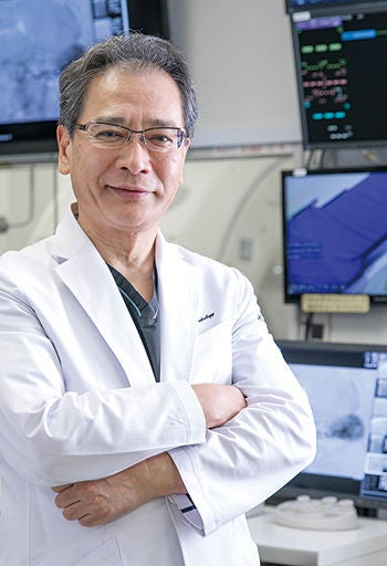
“The Alphenix 4D CT system is indispensable when performing extremely fine and selective arterial embolization, a method first introduced and currently being refined by Dr. Tanaka.”
Kimihiko Kichikawa, M.D.
Vice-President of Nara Medical University Hospital
Professor & Chairman of Dept. of Radiology
Director of Interventional Radiology Center
President of Japanese Society of Interventional Radiology (JSIR)
Q3: Have you seen a reduction in dose?
Dr. Tanaka: Compared to other conventional systems, the Alphenix 4D CT system significantly reduces the exposure dose to both patients and operators. With the SPOT ROI feature, the standard level of radiation exposure is used only in the area where precise visualization is required, and a lower level of radiation exposure is used in surrounding areas. As a result, the areas outside the ROI do not appear completely black but are shown with a certain degree of useful visual information. This allows the operator to perform procedures more comfortably.
In conventional procedures, we usually introduce the biopsy needle straight into the target under real-time CT fluoroscopic guidance. However, with Dynamic Volume CT, it is possible to slowly advance the needle while confirming the angle under MPR imaging based on the volume data, which minimizes the exposure dose (Direct MPR function).
Dr. Kichikawa: Radiation exposure has long been a challenge in CT-guided procedures, and the ability to perform such procedures in real-time without the need for fluoroscopy is, therefore, a significant benefit. In addition, the Dose Tracking System (DTS) of the angiography unit provides real-time exposure dose display, which helps limit the dose even further. When we perform angiography, we select the appropriate dose for the particular case and communicate with the technician to inform him or her whether precise visualization or only rough guidance is required. In addition, we are cautious to adjust the angle of the protective plate so that it is placed in proper contact with the patient. We also use goggles to protect the lenses of the eye from radiation exposure.
Q4: How can the Alphenix 4D CT system help with puncture procedures?
Dr. Kichikawa: The development of advanced puncture techniques has led to higher precision in puncture procedures. I expect this will also improve the accuracy of embolization for the management of endoleaks following stent placement in patients with aortic aneurysms.
Dr. Tanaka: I believe it will expand the range of possibilities in puncture procedures by allowing the needle to be introduced at oblique angles. In the conventional method, we could only introduce the needle in the x-axis or y-axis direction, but we can now insert the needle from a wide range of angles, making it easier to avoid sensitive anatomical structures. Previously, a puncture in the z-axis direction was only possible using the tilt method, and the angle was therefore limited to 30 degrees. Moreover, in vascular IR, the acquisition of 4D data is expected to expand the potential of this technique and to drive the development of new therapeutic methods in the future.
Q5: How would you like to see the Alphenix 4D CT system improved?
Dr. Kichikawa: Personally, I would wish for the availability of 3D fluoroscopy. Nowadays, we take 3D and 4D imaging for granted, but fluoroscopy is still limited to 2D.
Dr. Tanaka: From the viewpoint of puncture procedures, I would wish for faster real-time 3D reconstruction. I would also like to see advances in puncture angle guidance technology so that everyone can perform such procedures accurately and consistently.
Q6: What do you believe the future has in store for IR?
Dr. Kichikawa: Recently, there have been many reports on the use of IR in pain management. Percutaneous vertebroplasty has already been used to treat patients with vertebral compression fractures, and there have been some clinical trials involving the embolization of new blood vessels responsible for causing pain.
Dr. Tanaka: There have also been many studies conducted overseas to assess the potential efficacy of IR in patients with prostate cancer, in which extremely fine vessels need to be approached. I am extremely pleased to learn that the hybrid angiography-CT solution, which was first developed in Japan, is now widely recognized and is rapidly expanding worldwide.
Dr. Kichikawa: I believe it is inevitable that the hybrid angiography-CT solution will gain widespread acceptance in all parts of the world. This is because it is essential for taking IR to the next level. I look forward to continuing my work in order to further improve the system in collaboration with Canon Medical Systems and to expand the future potential of IR. //
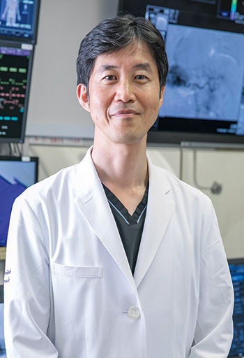
“The Alphenix 4D CT makes it possible to formulate precise therapeutic plans and to ensure the accurate implementation of IR.”
Toshihiro Tanaka, M.D., Ph.D.
Associate Professor
Dept. of Radiology, IVR Center
Social Media


