

Innovative High Definition (Hi-Def) Imaging System for Complex Interventional Cardiology Procedures
Andrew Kuhls-Gilcrist, PhD, DABR, Dale Marek, RT(R), Mark Hohn, Yiemeng Hoi, PhD
Medical Affairs, Interventional X-ray, Canon Medical Systems USA, Inc.
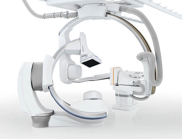
- Available as an option on Alphenix Core+ and Biplane systems with 12” detectors.
The new Alphenix family of interventional systems equipped with Hi-Def imaging* helps clinicians see fine details with clarity and precision during interventional procedures
Highest Resolution to Help Clinicians See Fine Details
The Alphenix interventional systems feature the all-new and exclusive high definition (Hi-Def) detector* with 76 micron pixel imaging modes, helping clinicians visualize anatomical details with clarity and deploy devices with confidence.
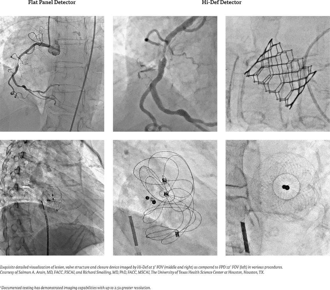
Featuring the world’s first Hi-Def detector – with more than twice the spatial resolution1 of conventional flat panel detectors (FPD) – for resolving fine details. This unique hybrid 12" x 12" FPD combines high definition imaging technology based on crystalline silicon that boosts spatial resolution up to 6.6 line pairs per millimeter.
Efficient & Seamless Workflow
The unique Alphenix system offers standard modes with 12", 10", 8", 6" or 4.3" fields of view (FOV) and three Hi-Def modes with 3", 2.3" or 1.5" FOV, delivering increased spatial resolution without interruption of procedure workflow. Alphenix’s advanced 16-bit imaging chain, including Illuvis triple-phase image processing and noise reduction technology, provides clean, sharp, more defined images during wire manipulation and device placement with enhanced visualization of the surrounding vessels and devices.
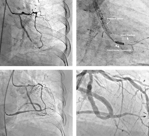 Occlusion of obtuse marginal artery of a previously placed stent imaged in 3" Hi-Def (top right) as compared to 8" FOV (top left) during a PCI procedure. The patient underwent successful angioplasty as shown in 3" Hi-Def (bottom right) as compared to 8" FOV at different angles (bottom left).
Case Report
Salman A. Arain, MD, FACC, FSCAI
and Richard Smalling, MD, PhD, FACC, MSCAI
The University of Texas Health Science Center at Houston, Houston, TX
History: A 61-year-old man with known coronary artery disease and prior stent placement within the native left circumflex artery system presented with unstable angina. Angiography showed occlusion of a previously placed stent in a large obtuse marginal artery.1
Procedure: Hi-Def imaging was utilized to enhance the visibility of the prior stent during PCI. FPD images were used for qualitative comparison. Radiation dose was measured using a real-time color-coded patient skin dose tracking system (DTS). The revolutionary DTS tracks and incorporates X-ray beam directions relative to the patient graphic, providing distribution of skin dose estimates rather than a single cumulative value. This exclusive monitoring technology has been shown to facilitate reduced radiation and helps to mitigate radiation risks.2,3
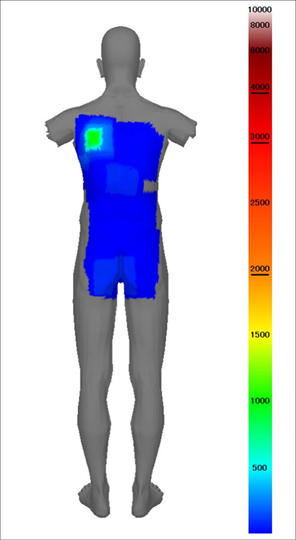 Real-time skin dose map generated by the dose tracking system (DTS) quantified a peak skin dose of only 1.0 Gy.
The patient underwent successful percutaneous coronary intervention using standard CTO techniques under Hi-Def guidance. The in-stent occlusion was crossed using a microcatheter and a 0.014 hydrophilic support wire. After successful angioplasty with a 2 mm balloon using standard magnification, high resolution imaging was used to study the stent architecture. Hi-Def imaging with a 3 inch FOV was able to clearly resolve individual stent struts and identify a stent fracture as well as under expanded stent not seen by standard FPD imaging. High pressure angioplasty was used to dilate the under expanded stent, and a 2.5 mm newer generation stent with thin struts was deployed within the obtuse marginal branch. During PCI, the DTS was leveraged to minimize radiation exposure. Despite irradiation and procedure times of 35 and 119 minutes, a peak skin dose of only 1.0 Gy was quantified, likely due to utilization of a smaller FOV over the treatment area in the Hi-Def mode. The patient made an uneventful recovery, and was free of angina at follow up.
Conclusion
Our initial experience using a novel Hi-Def imaging system shows that it can be safely used to improve visualization of coronary arteries and interventional devices during PCI in real-time with no observable increase in patient dose. Images obtained in the Hi-Def setting have a higher resolution and improved quality compared to standard imaging modes, allowing visual analysis of vessel anatomy and stent structure with a high level of accuracy.//
References
- S.A. Arain, et al., “Initial experience using a novel high definition (Hi-Def) imaging system in interventional cardiology,” J Am Coll Cardiol. 2020; Mar, 75 (11 Supplement 1) 3637.
- S.M. Wilson et al., “Real-time colour pictorial radiation monitoring during coronary angiography: effect on patient peak skin and total dose during coronary angiography,” EuroIntervention 2016; 12:e939-e947
- E. Ichimoto et al., “Efficacy of radiation dose reduction due to real-time monitoring and visualization of peak skin dose during coronary angiography and percutaneous coronary intervention,” Catheter Cardiovasc Interv. 2017; 1-6.
The clinical results, performance and views described in this case study are the experience of the author. Results may vary due to clinical setting, patient presentation and other factors. Many factors could cause the actual results and performance of Canon Medical’s product to be materially different from any of the aforementioned.
Social Media


