

Alphenix Biplane Hi-Def: A Point Where Cutting-Edge Technologies and Fine Art Meet
Dr. Ljubisa Borota, Uppsala University, Sweden
Canon Medical and the Department of Radiology, Uppsala University Hospital, Sweden, will celebrate this November the ten-year anniversary collaborative partnership in our neurointervention labora-tory. The relationship between Canon Medical and our section has never been an ordinary suppli-er-customer relationship. These ten years have been marked by mutual efforts in the development of the biplane system and on work that has focused on an analysis of the unique functionalities of this system. The conclusions of our strategic discussions are implemented by Canon’s R & D teams. All challenges have been overcome thanks to energy, motivation and, first of all, the deep mutual respect which was established during the many years of our collaboration. The Alphenix Biplane system that was launched was partly the result of these mutual efforts.
Our work was focused on an analysis of the unique functionalities of the Alphenix system that make it different and superior to other biplane systems.
The goal of our analyses was to become familiar with these novel functionalities in order to use them in our daily work in an optimal way. An additional objective was to publish our observations and the results of analyses in scientific journals. In this way the results of our work received scientific confirmation and became more accessible to practitioners and scientists in the field of neurointervention on a global level.
Dose saving functionalities
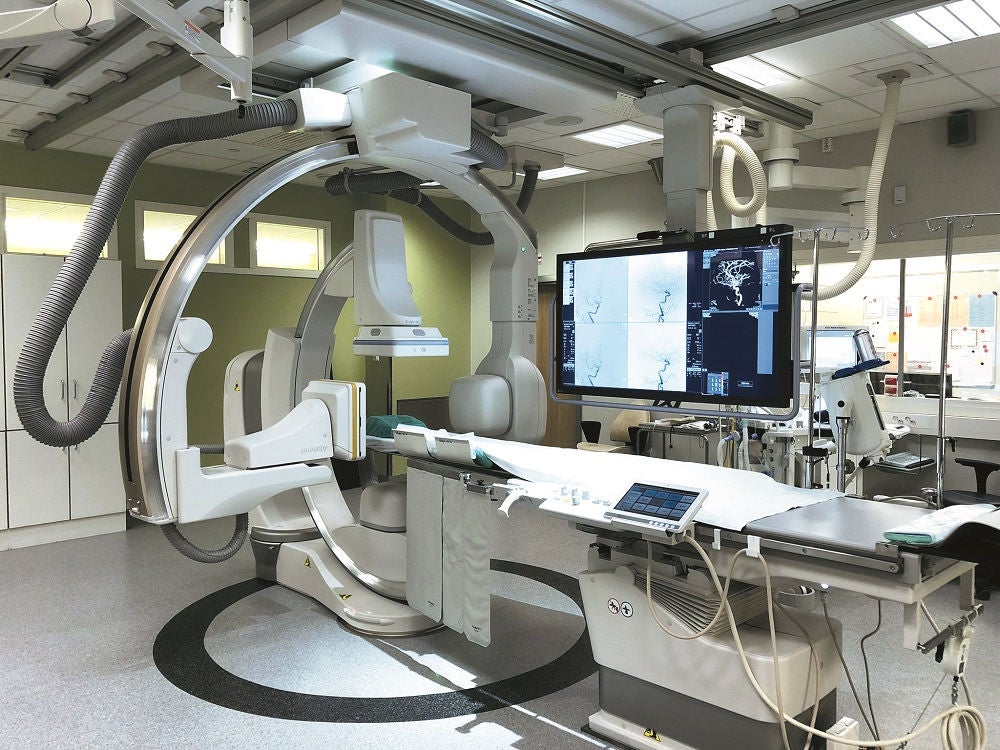 The Alphenix Biplane with Hi-Def at Uppsala University, Sweden.
Information regarding the dose delivered to the patient is of key importance to the operator. In all biplane systems except for the Alphenix Biplane, this information is shown in numerical form somewhere on the monitor together with less important information. Based on the research of Bednarek and colleagues1, Canon has developed a system that shows the area of the body that is exposed to the irradiation. The spectrum of colors in this area changes from dark blue to dark red, indicating delivered skin dose. This 3D model is displayed in the corner of the screen, and, since it is only a 3D color indicator, it keeps the operator continuously updated about the dose without being intrusive.
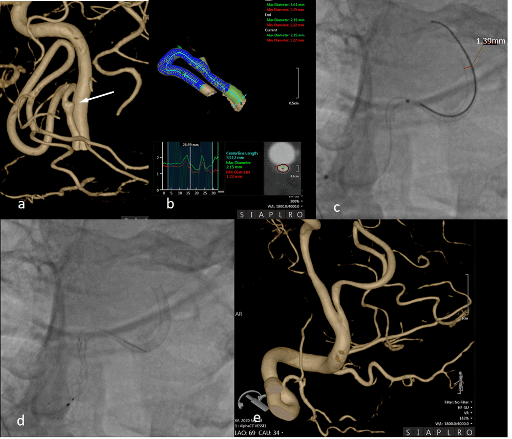
Figure 1:
- 3D reconstruction: dissecting aneurysm (arrow) of posterior inferior cerebellar artery (PICA).
- Virtual stenting indicates that the largest diameter of artery is 2.15 mm and the smallest 1.22 mm.
- High Def: a flow diverting stent is deployed.
- High Def: a second, braided stent is deployed in telescopic fashion in proximal segment of PICA and vertebral artery due to proximal extension of the dissection.
- 3D reconstruction: one week follow-up shows completely reconstructed artery.
Spot Fluoroscopy is a unique technology that enables acentric, asymmetric collimation anywhere within the field of view. The shape (square or rectangle), size or place of the region of interest defined by such collimation can be changed at any time during the intervention, and as many times as the operator needs to do it. Using this functionality, it is possible to adapt the field of view to the anatomy of a vascular target, enabling optimal visualization of the target with the lowest possible dose. We have shown that the dose delivered to a patient by using Spot Fluoroscopy is significantly lower than the dose delivered using conventional collimation2.
A similar, dose-sparing functionality referred to as Spot Region of Interest (Spot ROI), which is integrated in the Alphenix Biplane system, offers a square-shaped collimation of the region of interest that is freely movable within the field of view3. Unlike other collimation systems, the field of view outside the region of interest is still visible, which is particularly important in visualization of larger anatomical regions.
Another functionality that distinguishes this machine from any other biplane system is the flexible lateral isocenter. This functionality “enables mobility of the lateral arm in the vertical direction and in this way contributes to the adjustability of the lateral arm and thus to better visualization of vascular structures regardless of their position, size or shape.
Thanks to this novel functionality, it is also possible to obtain the desired projections with the minimal possible distance between the detector plate and X-ray tube.”4. A shorter source-detector distance also leads to a dose reduction according to the inverse square law. We have shown that the dose delivered to the target by using a flexible lateral isocenter is significantly lower than the dose delivered to the same target by using a fixed lateral isocenter4.
Beyond the imaginable…
Finally, the last jewel in the crown of Alphenix Biplane system is the High Definition (Hi-Def), one more functionality that distinguishes this machine from other biplane machines. The system is equipped with a novel hybrid flat panel detector on each plane that consists of a standard 30x30 detector and a built in 8x8cm High definition detector with a pixel size of 76µm providing 6,5LP/mm resolution. This unique high-resolution mode enables superior visualization of both the smallest anatomical structures and the smallest neurovascular devices. Clinical importance of this functionality has already been confirmed5. Manufacturing technology of neurovascular devices has developed tremendously during recent years. The range of sizes of the smallest aneurysms now treatable by endovascular means varies between 1 mm and 3 mm. The diameter of the loop of the smallest coil measures 1 mm and the total length of this coil measures 10 mm. High definition enables visualization of even the secondary structure of these coils.
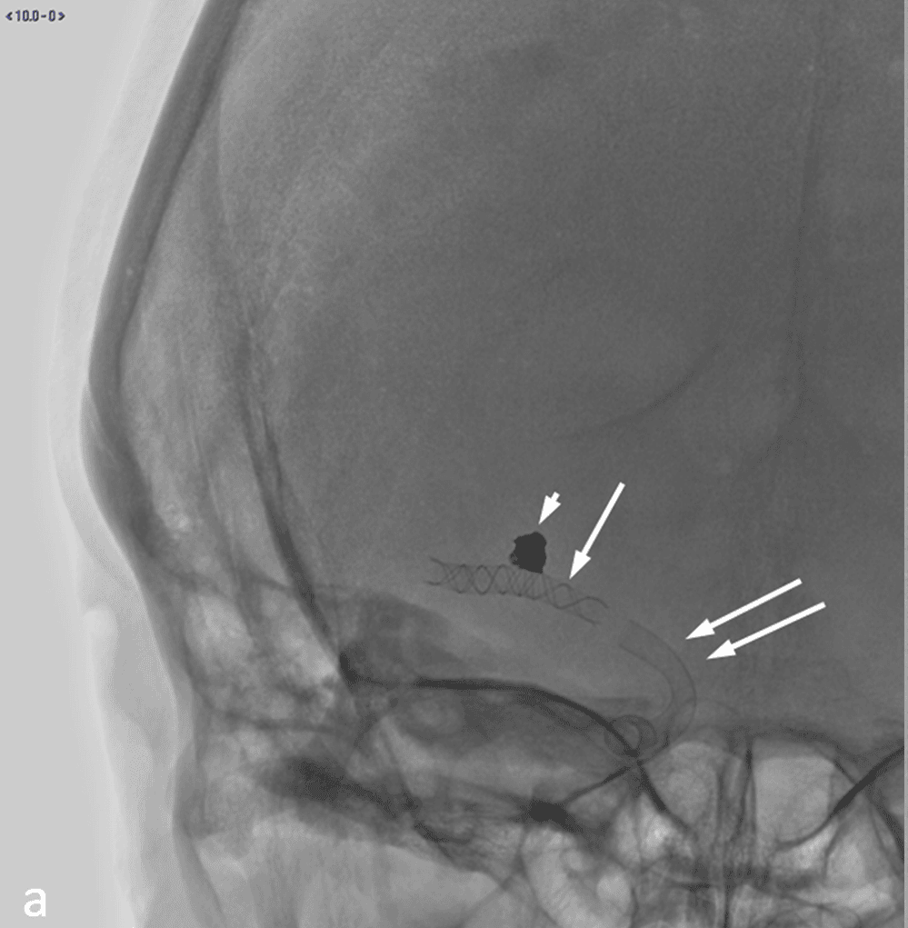
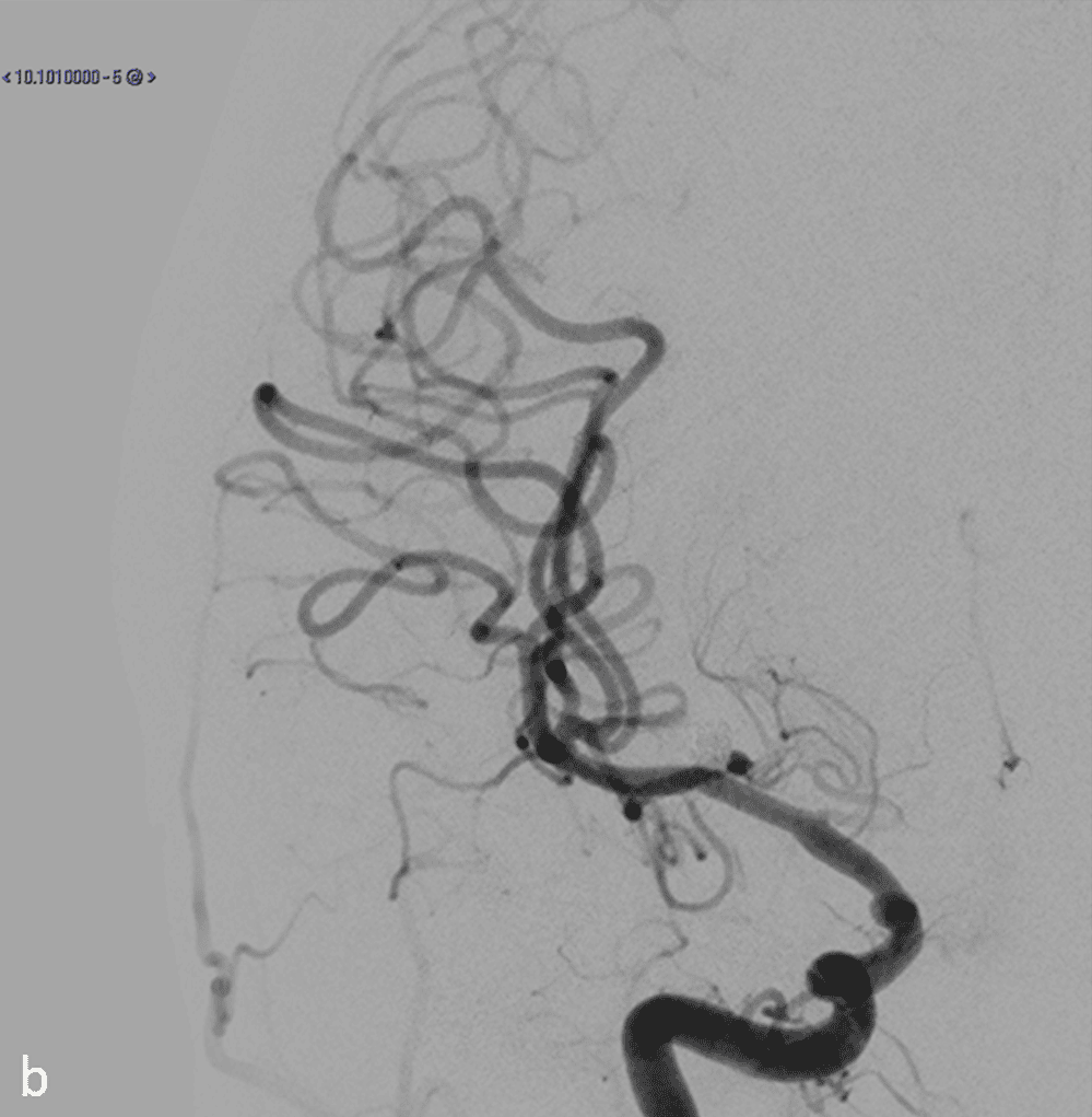
Figure 2:
- AP projection, spot image without magnification shows coils in an aneurysm (arrowhead), a flow diverting 2.5 x 25 Silk stent in middle cerebral artery (arrow) and a 3.75x25 Silk Vista Baby flow diverting stent in internal carotid artery (double arrow).
- Digital subtraction angiography, same projection without magnification.
Newly developed flow-diverting stents can be deployed even in arteries with a diameter between 1.5 mm and 2 mm (Figure 1). The new generation of these stents is fully visible thanks to new technologies that have enabled the manufacturing of the micro wires these stents are made of. Figures 2 and 3 illustrate in the best possible way the tremendous progress that has taken place in the development of endovascular devices. A wide-necked middle cerebral artery aneurysm was treated with coils and a flow-diverting stent in 2017. An ophthalmic artery aneurysm was treated with only a flow-diverting stent in 2021. Both stents were designed and produced by the same manufacturer. The image shown in the Figure 3 was taken using the High Definition function integrated into the Alphenix Biplane system. The first, strong impression is that the stent used in 2017 was not only partly visible but also primitive in comparison with the stent used in 2021. Even though the image is two-dimensional, it is not difficult to get a three-dimensional perception of a complex, tapered cylindrical structure made up of numerous densely braided micro wires and bent in several planes. The development of Canon’s Alphenix Biplane system very successfully follows the development of neurovascular devices. Thanks to this development, the treatment of even the smallest vascular targets with the finest neurovascular devices has never been as safe and reliable as it is now.
Instead of a conclusion...
 Dr. Ljubisa Borota, Uppsala University, Sweden.
The Alphenix Biplane is a unique system from several points of view. Elegantly designed arms combined with a flexible lateral isocenter offer almost unlimited adaptability to the shape and size of vascular targets. It is not an exaggeration to claim that impossible working projections do not exist with this machine. Multiple dose-saving functionalities contribute to the reduction of the dose to the patient and to the staff to the lowest possible values.
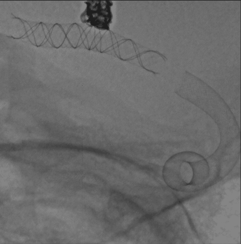
Figure 3:
The same case as on the Figure 2. Stents and coils are visualised by High Definition.
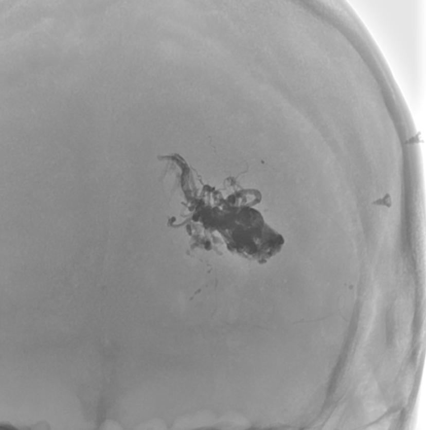
Figure 4:
Onyx cast in cerebral AVM … or in art analogy: the take-off of a black swan.
Neurointervention is somewhere between fine art and medicine… Sometimes, closer to fine art… The aim of our interventions is to re-open or reconstruct pathways for the blood supplying the brain or to fill bizarre cavities, aneurysm, fistulas or AVMs, with some of the numerous embolic materials available. A neurointerventionalist is like a sculptor who creates bizarre casts in the brain vasculature that remind one of modern abstract sculptures (Figure 4).
The High Definition function offers not only excellent visualization of the smallest anatomical structures and the smallest devices and their ultrastructure, but also offers the operator a unique visual, artistic experience. This machine is really the point where cutting-edge technologies and fine art meet. //
References
- Bednarek, Daniel, Barbarits, Jeffery, Rana, Vijay, Nagaraja, Srikanta, Josan, Madhur: Verification of the performance accuracy of a real-time skin-dose tracking system for interventional fluoroscopic procedures. Proc. SPIE 7961, Medical Imaging 2011: Physics of Medical Imaging, 796127 (16 March 2011); doi: 10.1117/12.877677
- Borota L, Jangland L, Åslund P-E, Ronne-Engström E, Nyberg C, Mahmoud E, Sakaguchi T and Patz A: Spot fluoroscopy: a novel innovative approach to reduce radiation dose in neurointerventional procedures. Acta Radiologica, 2017 May;58(5):600-608. doi: 10.1177/0284185116658682.
- Borota L, Patz A: Spot region of interest imaging: a novel functionality aimed at x-ray dose reduction in neurointerventional procedures. Radiat Prot Dosimetry, 2020 Jun 24;188(3):322-331. doi: 10.1093/rpd/ncz290.
- Borota L, Patz A: Flexible lateral isocenter: A novel mechanical functionality contributing to dose reduction in neurointerventional procedures. Interv Neuroradiol 2017 Dec;23(6):669-675. doi: 10.1177/1591019917728260.
- Nagesh SVS, Vakharia K, Waqas M, Munich S, Bednarek B, Davies JM , Kenneth V Snyder KV, Mokin M, Rudin S, Levy E, Siddiqui AH: Single-center experience of using high definition (Hi- Def) imaging during neurointervention treatment of intracranial aneurysms using flow diverters. J NeuroIntervent Surg 2020; 12:897–901. doi: 10.1136/neurintsurg-2019-015551
Visit our website:
https://eu.medical.canon/products/angiography/alphenix/neurology
Social Media


