

Experience using the New Alphenix Interventional System for Neuro-endovascular Therapy
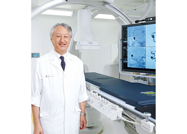
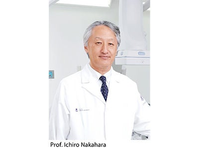
Prof. Ichiro Nakahara,
Department of Comprehensive Strokology,
Fujita Health University Hospital
Department of Comprehensive Strokology,
Fujita Health University Hospital
Critical Requirements for an Angiography System
Our most important requirement in considering a new angiography system is the ability to clearly visualize vessels and the tissues around vessels in both fluoroscopy and radiography. In addition, a sufficient range of functions are needed to support effective treatment, such as the growing importance of catheter-based procedures in current stroke care. Specifically, for 3D Angiography and the associated integrated workstations, as well as the various software applications supporting catheter-based procedures are considered essential.
However, we previously have encountered cases in which the conventional angiography system was unable to provide adequate fluoroscopic, radiographic, or 3D images of sufficient high quality. For example, halation has been often observed near the area where a direct X-ray beam enters, making it difficult to assess accurate information concerning the extracranial vessels and skull base. We also felt that there was a need for further improvement in the functionality of cone beam CT technology.
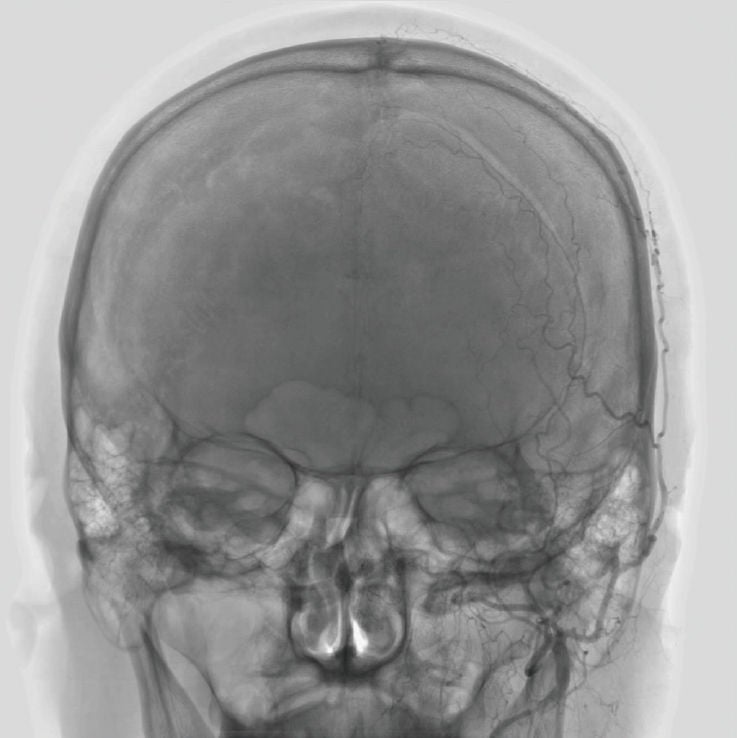
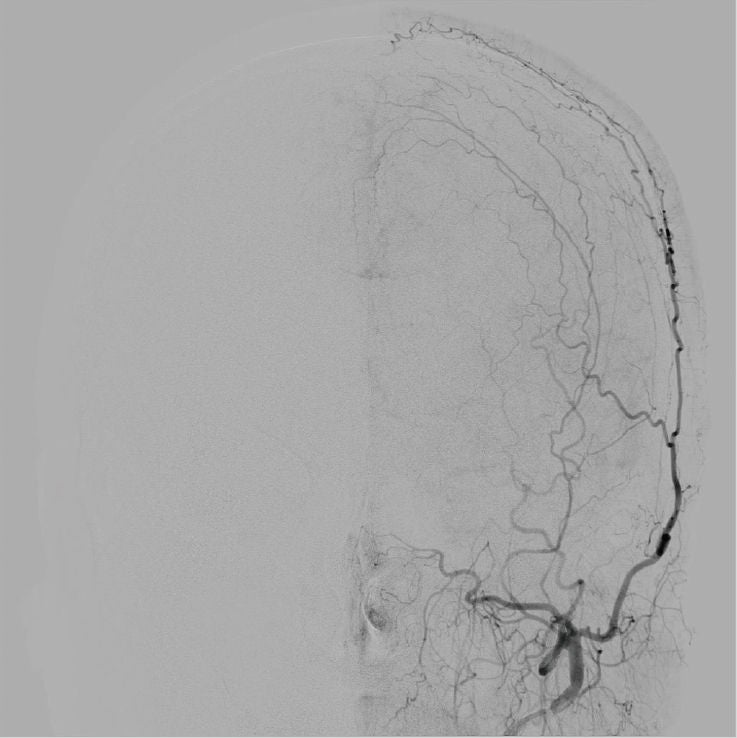
External Carotid Artery images.
Halation is not observed near the area where a direct X-ray beam enters, even without a compensation filter.
“During neurovascular therapy being able to see the intricate details in anatomy, and access devices gives us the confi dence to quickly and better treat our patients with less chance of complications.”
Features of the Alphenix System
The Alphenix system has been introduced to the market as a new angiography system driven by clinical needs and a development process focused on addressing and overcoming the above mentioned clinical limitations. Now with the detailed imaging information being provided, the necessary preciseness for proper clinical treatment of stroke can easily be attained. We are strongly confident that the Alphenix system can be utilized for all varieties of neuroendovascular procedures at a wide range of medical institutions around the world.
(1) Fluoroscopy/Radiography
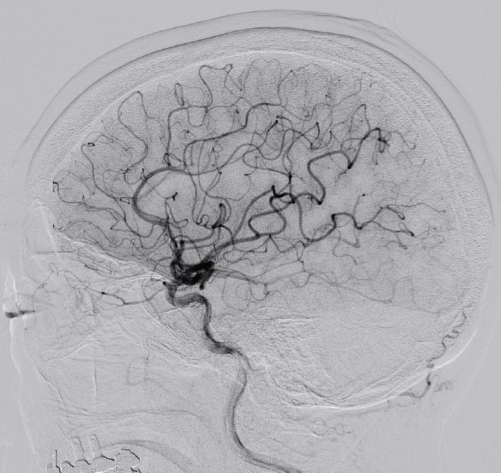
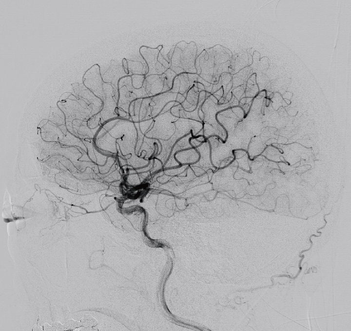
When using fluoroscopy on the Alphenix system, you notice the enhanced contrast in fluoroscopic images of the skull base. This makes it easier to clearly observe the precise movements needed to gain catheter access to tortuous vessel anatomy, the treatment zone, and the deployment behavior of the stent. Being able to visualize each platinum marker during the intracranial placement of flow-diverter stents can also be performed with greater confidence and ease. One of the useful features of this system is the advanced Auto-Pixel Shift function. The system instantly detects even the slightest patient movements and performs real-time pixel shifting to provide optimized subtraction images. This eliminates the time needed for the technologist to manually adjust the images, allowing the operating staff to focus on patient care, minimize surgical time, reduce complications, or to prepare for the next procedure immediately after the acquisition is completed.
Effects of Realtime Auto Pixel Shift
(2) 3D / Cone-Beam CT
Cone-beam CT technology plays a very important role in current neuroendovascular therapy. There is great need for a sophisticated cone-beam CT system with smart features for confirming precise stent placement and vessel wall apposition, as well as for observing fine perforator vessel anatomy.
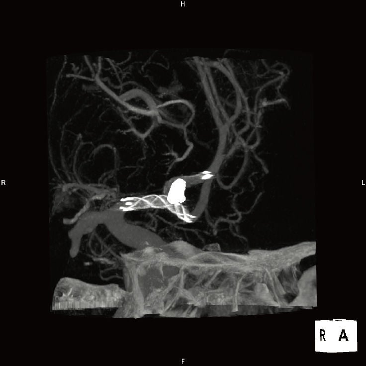
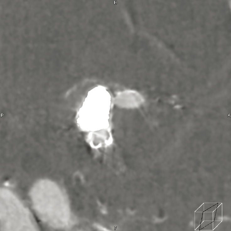
The current functional capabilities of cone-beam CT in conventional systems are considered inadequate for confirming the absence of intracranial hemorrhages during post treatment. However, thanks to the enhanced diagnostic performance of the Alphenix system, the presence or absence of a intracranial hemorrhage after treatment can be determined using our cone-beam CT function known as Alpha CT, which is currently in use at our facility. The most remarkable feature of Alpha CT is its capability to eliminate metal artifacts. When imaging a vessel in which endovascular coils have been placed, the artifacts caused by the coil elements can be eliminated, allowing the deployed stent placed in the vessel to be clearly visualized. In addition, images are generated at a higher speed, making it conveniently possible to quickly determine the current treatment status and proceed to the next step of the therapeutic procedure.
Alpha CT reduces Metal artifact
(3)Flexible Operability
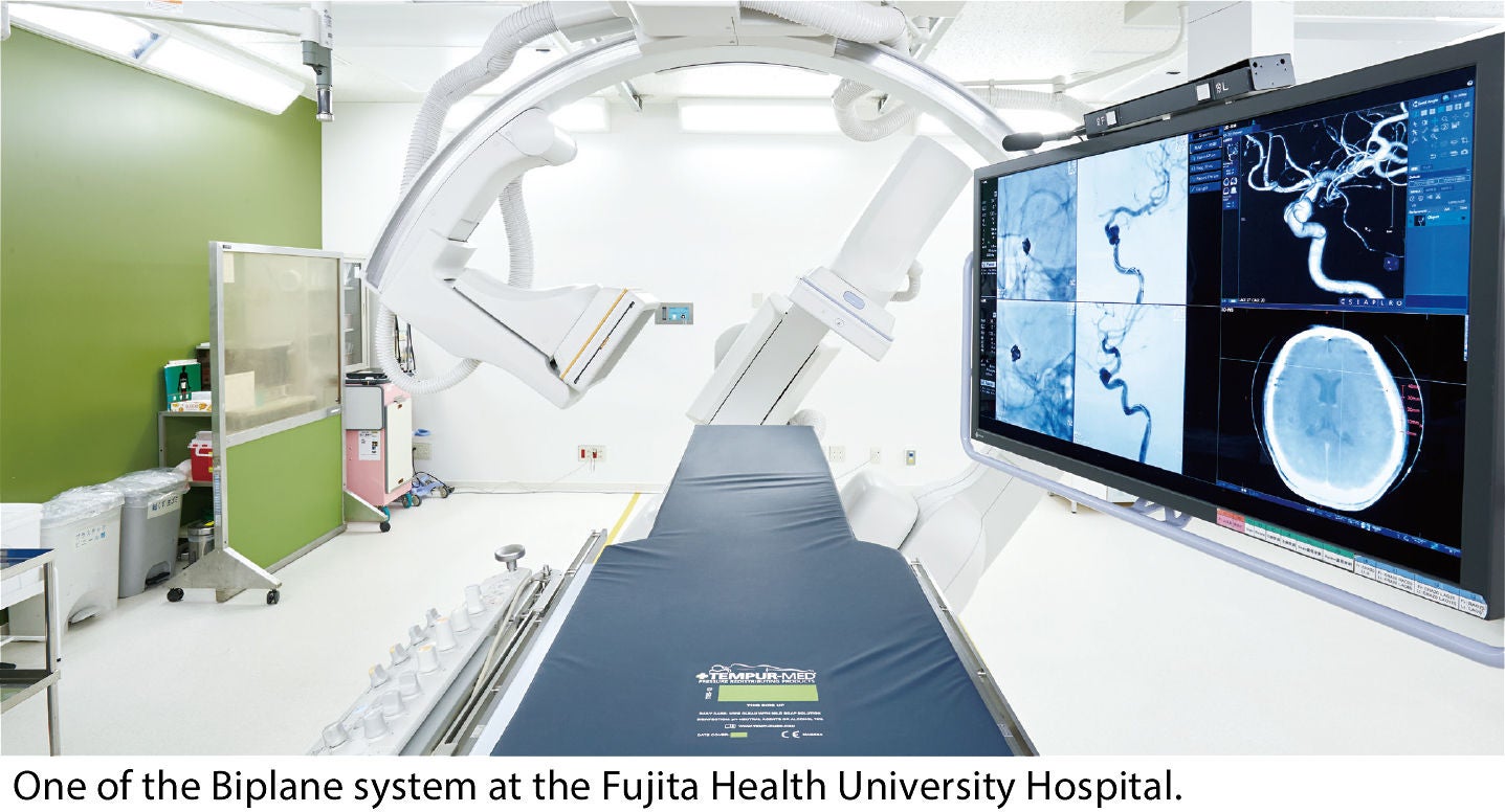
In neuroendovascular procedures, it is very important to set the appropriate working angle and to make use of the roadmapping function. In the Alphenix system, the working angle can easily be set from the 3D image in a single operation. In addition, a roadmap overlay can be generated instantly, as well a function for automatically generating the optimal mask image is also available. These functions are extremely effective, significantly reducing the clinical case workload on the operator as well as the time required for the therapeutic procedure.
However, now having experienced the Alphenix system in our clinical neuroendovascular treatment cases, it has since become our first choice. I have come to appreciate the fully-featured effectiveness of the Alphenix system, and I believe the same would be true for many other physicians who perform neuroendovascular procedures. Given the great technological advances in neuroendovascular therapy and the intricate devices, the requirements for an angiography systems technology must also evolve.
Angiography systems are not limited to use in the field of neurosurgery, but are often used in the field of cardiology and interventional radiology as well. We hope that angiography systems will continue to clinically evolve in parallel with the future progress of neurosurgical treatment and neuroendovascular procedures. Throughout the technical development process of the Alphenix system, I have contributed my clinical feedback and unbiased suggestions to help make critical feature improvements to the previous conventional system. The Canon Medical development and clinical applications teams took my feedback and suggested improvements from a clinical operator perspective very seriously. I believe that their sincere and responsive attitude has led to the developmental success of such a new intuitively dynamic and robust system.
Social Media


